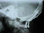
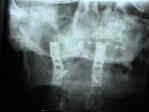
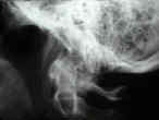
- See: cervical spine in RA
- Discussion:
- vertical subluxation is one of the most serious complications of rheumatoid arthritis, and may affect up to 5-10% of patients who
have cercial spine disease;
- basilar invagination may result from erosion and/or bone loss between occiput and dens or may result from erosion and settling
of the C1 / C2 articulation (? more common);;
- lateral masses of atlas may collapse secondary to erosive changes in atlantooccipital and atlantoaxial synovial joints;
- this leads to vertical sublux of odontoid process thru foramen magnum;
- tip of odontoid process, which may be expanded by surrounding pannus, is brought into contact with the cervicomedullary junction;
- SSEP may be helpful in evaluation;
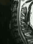
- Clinical Findings:
- need to rule out myelopathy:
- patients may note headaches due to compression of the greater occipital branch of C2;
- loss of pain / touch sensation over the trigeminal nerve distribution;
- sleep apnea;
- down beat nystagmus and inter-nuclear ophthalmoplegia;
- facial diplegia;
- dysphagia (due to CN IX dysfunction);
- Radiographic Work Up;
- Cross Table Lateral:
- in the report by Riew, et al. (2001), the authors attempted to validate and compare the most widely accepted plain radiographic criteria
for basilar invagination in this patient population;
- cervical radiographs of 131 rheumatoid patients were examined;
- 67 (29 with basilar invagination and 38 without it) were also evaluated with tomograms, MRI, and/or sagittally reconstructed
computed tomography scans to detect the presence of basilar invagination;
- radiographic criteria proposed by Clark et al., McRae and Barnum, Chamberlain, McGregor, Redlund-Johnell and Pettersson,
Ranawat et al., Fischgold and Metzger, and Wackenheim;
- no single test had a sensitivity and a negative predictive value of greater than 90% as well as a reasonable specificity and a
reasonable positive predictive value;
- combination of the Clark station, the Redlund-Johnell criterion, and the Ranawat criterion, scored as positive for basilar
invagination if any of the three were positive, proved to be better than any single criterion; the sensitivity of the combined
criteria was 94%, and the negative predictive value was 91%.
- ref: Diagnosing Basilar Invagination in the Rheumatoid Patient. The Reliability of Radiographic Criteria
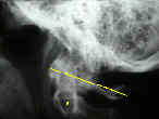
- Ranawat's line:
- center of C2 pedicle to a line connecting the anterior and posterior C1 arches;
- normal measurement in men is 17 mm, whereas in women it is 15 mm;
- distance of < 13 mm is consistent with impaction;
- less than 7 mm is associated with medullary compression on MR image
- McRae's line:
- defines the opening of the foramen magnum;
- the tip of the dens may protrude slightly above this line, but if the dens is below this line then impaction is not present;
- McGregor's line:
- line drawn from the posterior edge of the hard palate to the caudal posterior occipit curve;
- cranial settling is present when the tip of dens is more than 4.5 mm above this line;
- this measurement can be difficult when there is dens errosion;
- problem w/ this measurement is that the hard palate position may vary w/ mid facial anomalies;
- Chamberlain's line:
- line from dorsal margin of hard palate to the posterior edge of the foramen magnum;
- this line is often hard to visualize on standard radiographs;
- if dens is > 6 mm above this line, consistent w/ impaction;
- Indications for Surgery:
- progressive cranial migration or neurologic compromise may require operative intervention (occiput to C2 fusion);
- Treatment:
- when brain stem compromise is sig, w/ functional impairment, transoral or anterior retropharangeal odontoid resection may be required;
- fusion of occiput & second cervical vertebra for superior migration of odontoid process;
- w/ subaxial subluxation, perform posterior fusion & include occitput w/ fusion if superior migration of odontoid process is demonstrated;
- failure to extend the fusion to include subaxial subluxation, may lead to progressive instability;
- references:
- Incidence of subaxial subluxation in patients with generalized rhuematoid arthritis who have had previous ooccipital cervical fusions.
- resection of the odontoid:
- may be indicated for progressive neurologic involvement or cranial nerve involvement
Upper cervical instability in rheumatoid arthritis. Cranial subluxation of the odontoid process in rheumatoid arthritis. 3- to 11-year followup of occipitocervical fusion for rheumatoid arthritis.
Arthrodesis of the cervical spine in rheumatoid patients
The significance of certain measurements of the skull in the diagnosis of basilar impression.
Cervical spine fusion in rheumatoid arthritis
Fischgold H, Metzger J. Etude radiotomographique de l’impression basilaire. Rev Rhum Ed Fr. 1952;19:261.
Wackenheim A. Roentgen diagnosis of the cranio-vertebral region. New York: Springer; 1974.


