

 - Discussion:
 - w/ ankle fractures that occur above the syndesmosis, the lateral and/or medial malleolus, should be fixed first;
- subsequently, look for objective evidence of syndesmotic widening;
- stabilization of syndesmosis may be achieved by placing one or two screws between tibia & fibula to hold syndesmosis in
position until syndesmotic ligament healing can occur;
- syndesmotic screw is a positioning screw that is used to hold but not compress the syndesmosis;
- it is sometimes necessary to position the screw thru the fibular plate;
- in these cases, place plate along posterolateral fibular border, inorder to facilitate entry of syndesmotic screw into tibia;
- ref: Ankle fracture syndesmosis fixation and management: the current practice of orthopedic surgeons.
- Preoperative Planning:
- flouroscopy is required inorder to ensure that the screws are inserted at the proper level and are placed parallel to the joint line;
- if a lateral incision is require to fix a concomitant lateral malleolar frx, then place the incision more posteriorly inorder to facilitate screw insertion;
- patients need to be warned that hardware failure (screw breakage) is a common complication and does not imply that there was a surgical error;
- Weber C Fractures:
- Clinical use of a syndesmosis screw in stage IV pronation-external rotation ankle fractures.
- Ankle mortise stability in Weber C fractures: indications for syndesmotic fixation.
- The influence of a diastasis screw on the outcome of Weber type-C ankle fractures.
- Proper Level for Syndesmotic Screw:
- place the first screw approximately 1 cm proximal to syndesmosis or 4 cm proximal to the ankle joint;
- if it is too low, it may pass thru the distal tibio-fibular articulation (or may pass thru interosseous ligament) which can localized calcification and/or pain;
- if the screw insertion is too high, it may cause the tip of the fibula to toe outward;
- it is also important to direct the screws parallel to the joint line inorder to avoid tilting the distal fibula;
- following insertion, get films to r/o malreduction of the fibula in notch of tibia, with inferior and anterior subluxation of talus;
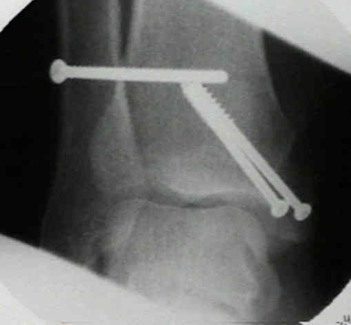
- in the following examples the syndesmotic screws have been placed too low which may lead to chronic syndesmotic pain (especially
if the screw breaks within the tibial-fibular joint);
- references:
- Syndesmotic screw placement: a biomechanical analysis.
- Technical aspects of the syndesmotic screw and their effect on functional outcome following acute distal tibiofibular syndesmosis injury.
- Biomechanical evaluation of syndesmotic screw position: a finite-element analysis.


- Hardware:
- number of screws and number of cortices:
- as noted by Mulligan and Hopkinson (Xenos, et al. 1995), repair with 2 screws inserted through 3 cortices each, proveded significantly
more stability than 1 screw;
- if screw engages only 3 cortices, normal external rotation of fibula during dorsiflexion of the ankle will not be affected;
- if adequate purchase is not achieved with 3 cortices, then far cortex should be fixed (4 cortices);
- if movement occurs, this screw will loosen in lateral cortex of tibia rather than break;
- screw engagement in the far cortex risks screw breakage;
- consider need for only one screw in Weber B fractures;
- references:
- The tibiofibular syndesmosis. Evaluation of the ligamentous structures, methods of fixation, and radiographic assessment.
- Tricortical Versus Quadricortical Syndesmosis Fixation in Ankle Fractures: A Prospective, Randomized Study Comparing Two Methods of Syndesmosis Fixation.
- Syndesmosis fixation: a comparison of three and four cortices of screw fixation without hardware removal.
- No difference in functional and radiographic results 8.4 years after quadricortical compared with tricortical syndesmosis fixation in ankle fractures.
- The management of acute distal tibio-fibular syndesmotic injuries: Results of a nationwide survey.
- Removal of the syndesmotic screw after the surgical treatment of a fracture of the ankle in adult patients does not affect one-year outcomes: a randomised controlled trial.
- size of screws:
- 3.5 mm: these should only be used in smaller patients;
- 4.5 mm screw (3.2 mm drill bit) usually 45 mm in length;
- in the abstract by Thompson MC, et al, the authors examined syndesmotic fixation with either 3.5 or 4.5 mm tricortical screws;
- the authors noted that both the 3.5 and 4.5 mm screws demonstrated similar biomechanical characteristics and did not find a
biomechanical advantage of a 4.5-mm screw over a 3.5-mm in fixation of the syndesmosis;
- references:
- Biomechanical comparison of syndesmosis fixation with 3.5- and 4.5-millimeter stainless steel screws.
- The fate of syndesmotic screws.
- Reduction of Syndesmosis
- Screw Insertion:
- prior to screw insertion, one assistant must firmly hold the fibula and the tibia together, so that the drill holes will not shift;
- screw direction:
- because fibula is posterior to tibia, syndesmotic screw must be angled from posterolateral to anteromedial in order to engage the tibia;
- if the drill is directed straight medially, it will skive off the posterior tibial surface;
- screw is inserted obliquely from back to front at angle 25 deg to 30 deg;
- it is directed from lateral to the medial cortex of the fibula, and into lateral cortex of the tibia at right angles to the long axis;
- it is essential that the screw also be directed parallel to the joint line;
- if the screw is directed proximally or distally there may be shortening or lengthening at the frx site;
- screw should be parallel to joint to avoid displacement of fibula in an inferior or superior direction (use flouroscopy);
- hardware:
- using the 2.5 mm drill bit in the double drill sleeve end, 2.5 mm both cortices of the fibula and at least one of the tibia;
- 3.5 mm tap is applied across the lateral tibial surface, while the fibula is held in firm reduction;
- this screw is placed either thru fibula or thru one of holes in fibular plate;
- fixation w/ lag screw is not performed since this could result in overtightening of screw and narrowing of syndesmosis;
- if this happens, dorsiflexion of ankle will be diminished;
- syndesmotic reduction:
- complications: Anterior & Posterior Subluxation of Fibula:
- this problem may be seen w/ improperly fixed syndesmotic injuries;
- this occurs when fibula is screwed to the tibia w/o visualization of the notch to ensure it is properly reduced;
- recognition that there is a problem is easy because the talus is subluxated in the same direction as the fibula is displaced;
- removing the bolt, repositioning the fibula in the notch under direct vision, and reinserting the bolt corrects the problem;
- PostOp Care:
- Wt Bearing:
- whether to allow wt bearing is controversial;
- concern is that screw will break w/ wt bearing;
- many advocate removal of syndesmotic screw before wt-bearing;
- if screw engages only 3 cortices, normal external rotation of fibula during dorsiflexion of the ankle will not be affected;
- radiolucency has been noted around such screws after pts have started to bear weight, suggesting that loosening of screw permits
normal motion of fibula;
- Screw Removal:
- many authors recommend removing the screw prior to full wt bearing, since the syndesmosis screw tends to limit ankle motion (which would
then transfer wt bearing forces against the screw - leading to breakage);
- generally syndesmotic screws are removed at 9-12 weeks, but some wait up to 4 months (to avoid late syndesmotic widening);
- bioabsorbable screw:
- at the time of screw removal, consider insertion of a bioabsorbable screw which will provide continued syndesmotic fixation,
and screw breakage is not an issue;
- references:
- The Radiographic Fate of the Syndesmosis after Trans-syndesmotic Screw Removal in Displaced Ankle Fractures
- Functional outcomes after syndesmotic screw fixation and removal.
- Functional and radiographic results of patients with syndesmotic screw fixation: implications for screw removal.
- To retain or remove the syndesmotic screw: a review of literature.
- Functional outcomes following syndesmotic fixation: A comparison of screws retained in situ versus routine removal - Is it really necessary?
Outcome after unstable ankle fracture: effect of syndesmotic stabilization.
Tibiotalar joint dynamics: indications for the syndesmotic screw--a cadaver study.
Mechanical considerations for the syndesmosis screw. A cadaver study.
The effect of the syndesmotic screw on the extension capacity of the ankle joint.
Stablization of ankle syndesmosis injuries with a syndesmosis screw.
- Anterior Syndesmosis;
- lag screw fixation if avulsed from bone (tubercle of Tillaux-Chaput), otherwise possible suture of ligament;
- if instability persists, 3.5 mm cortex screw is inserted as fibulo-tibial transfixation (positioning) screw about 2-3 cm above the ankle;
- screw is inserted obliquely 25-30 deg starting posterolaterally & aiming anteromedially;
- this screw is fully threaded, not a lag screw;
- bioabsorable screw fixation:
- in the report by Thordarson, et al., 32 patients who had pronation-lateral rotation (PLR) fractures occurring 4 centimeters or more proximal to
the ankle joint or lower if the talus was displaced greater than one centimeter laterally were enrolled in this study;
- 17 patients were randomized to fibular plate fixation with a 4.5 ml polylactic acid (PLA) bioabsorbable syndesmotic screw, and 15
patients randomized to fibular plate fixation with a 4.5 mm stainless steel syndesmotic screw;
- all 32 patients had uncomplicated healing of their fibular fracture without loss of reduction;
- there was neither evidence of osteolysis nor sterile effusion in the patients who were treated with the PLA screw;
- no difference in range of motion or subjective complaints was noted in either group;
- use of the PLA syndesmotic screw at short-term follow-up was well tolerated and avoided the need for subsequent screw removal;
- in the report by Hovis WD, et al (2002) the authors assessed the efficacy of screws made of polylevolactic acid (PLLA) in the treatment
of syndesmotic disruptions associated with ankle fractures and fracture-dislocations;
- 33 consecutive patients with a syndesmotic disruption were managed with standard metallic plate-and-screw fixation of the malleolar
fracture and with 4.5-mm polylevolactic acid screws, with purchase in four cortices, for fixation of the syndesmosis;
- all of the patients were managed with a non-weight-bearing plaster splint or brace for six weeks; or widening of the medial clear
space was detectable on radiographs;
- 19 patients (83%) had an excellent result, and four patients (17%) had a good result;
- no patient had malunion, nonunion, loss of reduction, or complications attributable to the biomechanical or biochemical properties of the implants;
- technical considerations:
- insert one 4.5 mm polylevolactic screw across syndesmosis with purchase obtained in 4 cortices approx 1-2 cm proximal to plafond;
- w/ Maisonneuve fractures, 2 screws were inserted;
- references:
- Bioabsorbable versus stainless steel screw fixation of the syndesmosis in pronation-lateral rotation ankle fractures: a prospective randomized trial.
- Treatment of Syndesmotic Disruptions of the Ankle with Bioabsorbable Screw Fixation