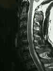
- Discussion:
- flexion-extension laterals;
- prior to intubation for elective surgery, all patients at risk should have flexion extension laterals of cervical spine r/o
atlantoaxial instability;
- Radiographs:
- atlantodens interval (ADI) needs to be evaluated;
- instability is present when a 3.5 mm ADI difference on flex/ext views;
- 7 mm difference may imply disruption of the alar ligaments;
- difference of > 9 mm is associated with an increase in neurologic injury and will usually require posterior fusion and wiring;
- space available for cord (SAC):
- posterior space of < 13 mm is contraindication to elective surgery until C-spine is stabilized first;
- Radiographic Evaluation:
- Cross Table Lateral:
- McGregor's line:
- hard palate-posteror occipit curve;
- dx is based on tip of dens being > 8 mm above this line in men and > 10 mm above the line in women;
- Ranawat's line:
- center of C2 pedicle to the C1 arch;
- nl is > 17 mm and < 13 mm is consistent with impaction;

- Chamberlain's line:
- anterior foramen to the top of the C1 arch;
- if the dens is > 6 mm above this line, consistent w/ impaction


