- See:
- Green Stick Fractures
- Pediatric Distal Radius Fracture
- Pediatric Ulnar Fracture
- Discussion:
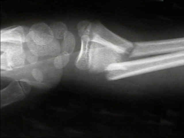
- frx may be of green stick type or complete (latter may be undisplaced, minimally displaced, or markedly displaced w/ overridding);
- frx may be greenstick or complete in both the radius and ulna, or it may be complete in one bone and green stick in the other;
- angulation may be volar, dorsal, or toward or away from interosseous space (see: deforming forces) 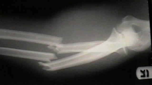
- mechanism:
- indirect injury during fall on an outstretched hand;
- direct violence occassionally is cause of both bone forearm frx;
- child abuse: in infants (less than one year), there is a high incidence of child abuse;
- frx location:
- proximal third fractures
- middle third:
- account for 18% of both bones fractures;
- these fracutres are often unstable and may be difficult to reduce with casting due to the thickness of the overlying muscle mass;
- displaced midshaft fractures w/ radial fracture proximal to the ulnar fracture are especially unstable, and are prone to redisplacement;
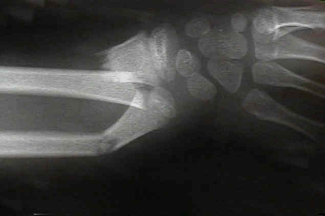
- distal third frx:
- account for 75% of frx of the shaft of the radius and ulna;
 *
*  *
* 
 *
*  *
* 
- Exam:
- evaluate AIN and PIN;
- note firmness of the volar and dorsal compartments;
- compartment syndrome;
- note that unlike adults, children may develop delayed compartment syndrome
- reference:
- Compartment syndrome following intramedullary fixation of pediatric forearm fractures.
- Acute Compartment Syndrome After Intramedullary Nailing of Isolated Radius and Ulna Fractures in Children
- Radiographs:
- radiographs from the wrist to the elbow (r/o radial head fracture)
- when only one bone of forearm is broken, the integrity of the proximal & distal RU joint should always be determined by obtaining x-rays that
include the elbow and wrist with the entire forearm;
- rotational alignment:
- normal, uninjured radius: the bicipital tuberosity is 180% to the radial styloid;
- look at the differences in cortical widths of frx edges;
- relation of bicipital tuberosity to radial styloid;
- in full supination bicipital tuberosity should be prominent on ulnar side of radius;
- in neutral rotation the bicipital tuberosity should not be seen;
- rotational alignment of proximal fragment:
- bicipital tuberosity is the landmark for identifying the rotational position of the proximal fragment;
- 90 degrees of supination: It is directed medially.
- neutral: It is directed posteriorly.
- 90 degrees of pronation: This is directed laterally.
- Acceptable Reduction: (Reduction of Both Bone Forearm Fractures):
- common radiographic pitfalls:
- the radiograph may not have been taken in the plane of maximal deformity, and therefore, x-rays will falsely minimize the degree of deformity;
- do not assume that the fracture position is static, ie, a fracture may show an "acceptable 15 deg" deformity at day 10, only to increase to 20 deg on day 20 (at
which point closed reduction may not be possible);
- Closed Treatment
- refernces:
- Risk Factors Associated with Loss of Position After Closed Reduction of Distal Radial Fractures in Children
- Re-displacement of stable distal both-bone forearm fractures in children: A randomised controlled multicentre trial.
- Operative Treatment: (see synthese technique manual)
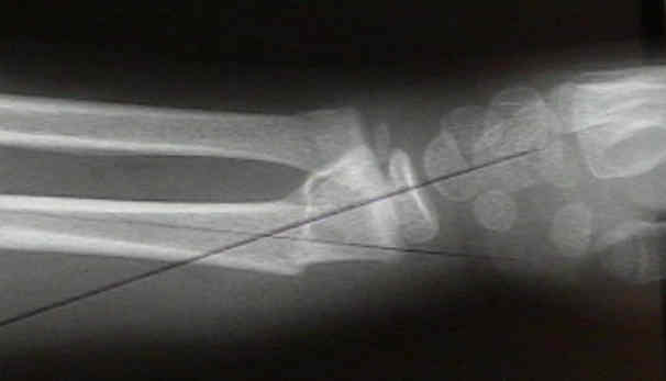
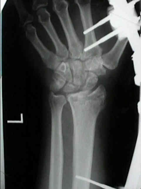
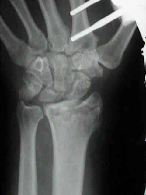
- straight K wire fixation for distal fractures;
- one K wire is inserted thru the frx site and is levered to achieve the reduction;


- Flexible intramedullary fixation:
- attractive in children because an incision is avoided, but the complication rate is high;
- limited open incisions and cross pinning allows direct anatomic fixation of the fractures and reliable healing;
- in the report by Houshian, et al., nail removal was undertaken at a median of 19 weeks (range 16-24 weeks) post-operatively;
- technique pearls:
- rather than using the traditional radial styloid approach, consider nail insertion in the ulnar aspect of the radius (proximal to physis);
- consider the more optimal reduction and the restoration of the radial bow, as compared to the photo out of the Synthes technique manual (right)
- Forearm fractures in children. Single bone fixation with elastic stable intramedullary nailing in 20 cases.
- references:
- Closing intramedullary nailing for the treatment of diaphyseal forearm fractures in adolescence: a preliminary report.
- Elastic stable intramedullary nailing in forearm shaft fractures in children: 85 cases.
- Use of pins and plaster in the treatment of unstable pediatric forearm fractures.
- Flexible intramedullary nailing as fracture treatment in children.
- Shaft forearm fractures in children: intramedullary nailing with immediate motion. A preliminary report.
- Open reduction and internal fixation of forearm fractures in children.
- Open reduction and internal fixation of pediatric forearm fractures. 
- Forearm fractures in children. Single bone fixation with elastic stable intramedullary nailing in 20 cases.
- Treatment of unstable fractures of the forearm in children. Is plating of a single bone adequate?
- Complications and outcomes of open pediatric forearm fractures.
- Percutaneous transphyseal intramedullary Kirschner wire pinning: a safe procedure for treatment of diaphyseal forearm fracture in children.
- Outcomes of intramedullary nail fixation through the olecranon apophysis in skeletally immature forearm fractures.
- Flexible intramedullary nailing of displaced diaphyseal forearm fractures in children.
- Use and abuse of flexible intramedullary nailing in children and adolescents.
- Comparison of Intramedullary Nailing to Plating for Both-Bone Forearm Fractures in Older Children.
- Use of Elastic Stable Intramedullary Nailing for Treating Unstable Forearm Fractures in Children.
- Eleven Years Experience in the Operative Management of Pediatric Forearm Fractures
- Failures and complications in intramedullary nailing of children's forearm fractures
- Study: Avoid elastic IM nailing in pediatric radius fractures with small distal fragments
- Single-bone intramedullary fixation of unstable both-bone diaphyseal forearm fractures in children leads to increased re-displacement: a multicentre randomised controlled trial.
- Complications and Outcomes of Diaphyseal Forearm Fracture Intramedullary Nailing: A Comparison of Pediatric and Adolescent Age Groups
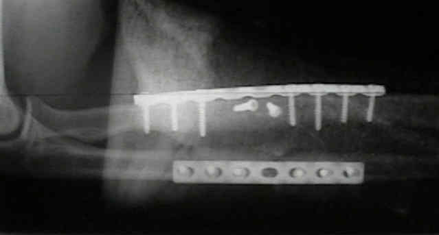
- plate fixation of pediatric forearm fractures: (see adult forearm frx)
- in the report by Bhaskar, et al, authors studied, retrospectively, 32 unstable fractures of forearm in children treated by compression plating;
- group A (20 children) had conventional plating of both forearm bones and group B (12 children) had plating of the ulna only;
- in group B an acceptable position of the radius was regarded as less than 10° of angulation in both AP and lateral planes, and with bone ends hitched;
- this was achieved by closed means in all except two cases, which were therefore included in group A;
- union was achieved in all patients, the mean time being 9.8 weeks in group A and 11.5 weeks in B;
- after a mean interval of at least 12 months, 14 children in group A and 9 in group B had their fixation devices removed;
- in group A, complications were noted in 8 patients (40%) after fixation and in six (42%) in relation to removal of the radial plate;
- no complications occurred in group B;
- final outcome for 23 patients was excellent or good in 12 of 14 (90%) in group A, despite the complications, and in eight of nine in group B (90%).
- authors recommend that if reduction and fixation of the fracture of the ulna alone restores acceptable alignment of the radius in unstable fractures
of the forearm, operation on the radius can be avoided;
- references:
- Treatment of unstable fractures of the forearm in children. Is plating of a single bone adequate?
- Unstable diaphyseal fractures of both bones of the forearm in children: plate fixation versus intramedullary nailing.
- Flexible intramedullary nailing of displaced diaphyseal forearm fractures in children.
- Complications:
- compartment syndrome:
- note that many forearm compartment syndromes will occur during the postoperative period (as opposed to preop_
- references:
- Complications and outcomes of diaphyseal forearm fracture intramedullary nailing: a comparison of pediatric and adolescent age groups.
- Acute Compartment Syndrome After Intramedullary Nailing of Isolated Radius and Ulna Fractures in Children
- neurologic injury:
- median, PIN, and ulnar nerve injuries;
- reference:
- Combined entrapment of the median and anterior interosseous nerves in a pediatric both-bone forearm fracture.
- malunion:
- Outcome after corrective osteotomy for malunited fractures of the forearm sustained in childhood.
- Malunited fractures of the forearm in children.
- Malunited forearm fractures in children.
- refracture: most common is first 6 mo.
- The Healing Forearm Fracture: a Matched Comparison of Forearm Refractures.
- Redisplacement of Diaphyseal Fractures of the Forearm After Closed Reduction in Children: a Retrospective Analysis of Risk Factors
- tendon rupture:
- Extensor pollicis longus rupture after fixation of radius and ulna fracture with titanium elastic nail (TEN) in a Child: a case report.
- synostosis: rare:
- risk factors:
- displaced frx w/ elevated thick periosteum;
- severe/surgical trauma (esp if treated w/ one incision)
- repeated manipulations;
- excision of the cross-union does not work as well in children as adults;
- in the report by Aner A, et al, the authors discuss the interposition of Gore-Tex vascular graft material between the synostosis;
- reference:
- Cross-union complicating fracture of the forearm. Part II: Children.
- Posttraumatic radioulnar synostosis treated with a free vascularized fat transplant and dynamic splint: a report of two cases.
- Surgical Treatment of Posttraumatic Radioulnar Synostosis in Children
- References:
- Displaced diaphyseal forearm fractures in children: classification and evaluation of the early radiographic prognosis.
- Pattern of forearm fractures in children.
- Forearm fractures in children. Pitfalls and complications.
- More Case Examples: