

- Indications for ORIF:
- failure of closed reduction and pecutaneous pin fixation;
- fractures presenting w/ more than than 2 mm of displacement and greater than 15 deg of talometatarsal angulation);
- young competitive atheletes may require anatomic reduction;
- disrupted skin and excessive swelling are relative contra-indications for ORIF;
- Technique:
- exposure:
- reduction:
- open the capsule of the 2nd MT-middle cuneiform to expose the joint surfaces;
- implants: 3.5 mm or 4.5 mm screws (either cortical or cannulated);
- fixation statedgy:
- medial-middle cuneiform articulation:
- if it is displaced, it is reduced and held w/ K wires;
- 1st metatarsal - medial cunieform articulation:
- ensure that navicular-cuneiform complex is intact;
- ensure that the 1st metatarsal is plantar flexed to an appropriate degree (this is often difficult to judge);
- 1st TMT joint was aligned by opposing medial border of medial cuneiform to the medial border of 1st metatarsal;
- plantar-medial aspect of this joint needs to be visualized and assurance made that there is no plantar gap;
- notch is made in dorsal cortex of proximal shaft of 1st metatarsal about 1.5 to 2 cm distal to the joint;
- it minimizes prominence of the screw head under the skin and prevents screw head from causing frx displacement;
- K wire is inserted from upper edge of notch across base of first cuneiform, aiming in a slight plantar direction;
- the cannulated screw is not placed until the other joint are reduced w/ K wires (to ensure that screws will not impinge on each other);
- in male pts w/ large 1st metatarsal shaft, use 4.0 to 4.5 mm cannulated screws (or alternatively use, 3.5 mm cortical lag screw);
- screw length is typically 35 to 40 mm;
- 2nd metatarsal-medial cuneiform (lisfranc's complex);
- reduction of frx dislocation of 2nd metatarsal is essential, but fixation is optional especially if 2nd metatarsal is fractured;
- one option is stabilization of 2nd metatarsal base to middle cuneiform w/ a 4.0 cannulated screw;
- lisfranc screw::
- insertion of cannulated screw (4.0 mm) from medial cuneiform into base of second metatarsal;
- increases the stability of the fixation;
- 3rd metatarsal:
- if base of 2nd metatarsal is frx'ed, procede to reduction of 3rd cuneiform joint;
- in some cases a second dorsal incision is required for this reduction between the 3rd and 4th metatarsals to expose the 3rd metatarsocuneiform joint;
- 4th and 5th metatarsal - cuboid joints:
- in most cases, reduction of the medial three rays will tend to reduce the 4th and 5th metatarsals;
- these metatarsals have more mobility that medial ones and therefored, rigid fixation is more likely to results in symptomatic stiffness;
- hence these joints may be best fixed with K wires rather than screws;
- further, the second incision, risks wound slough;
- if possible consider use of percutenous pins for fixation, from bases of 4th and 5th metatarsal shafts into the cuboid;
- if percuneous pins are not considered adequate, then consider percutaneous screw insertion through the metatarsal shaft, into the cuboid;
- Complications:
- broken hardware: screws may break in up to 25% of patients;
- degenerative arthritis: may be seen in upto 25% of patients

Outcome After Open Reduction and Internal Fixation of Lisfranc Joint Injuries
Intramedullary transmetatarsal Kirschner wire fixation of Lisfranc fracture-dislocations.
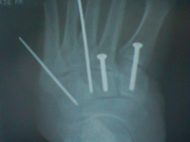
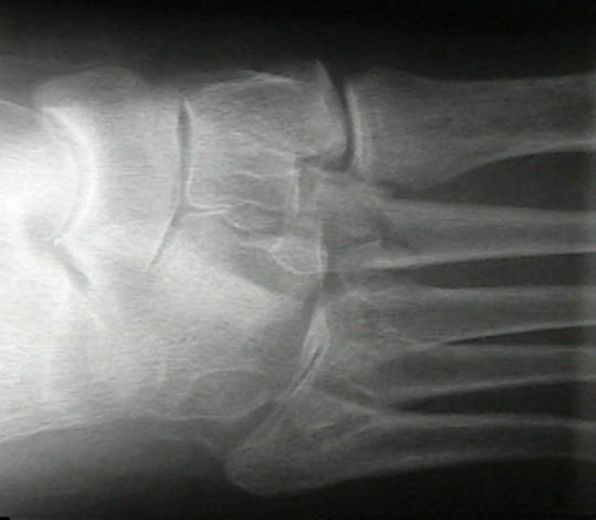
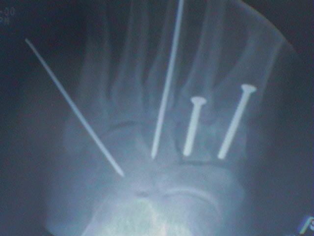
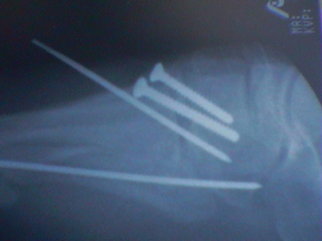
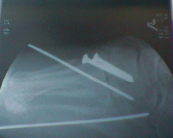
- Case Examples:


