

- See: Midfoot/Forefoot Fractures
- Discussion:
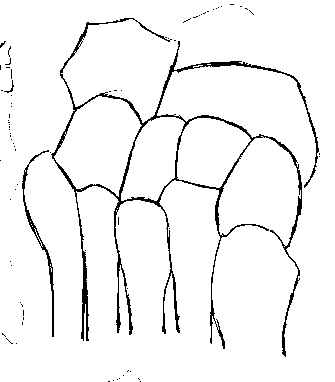
- anatomy of the midfoot
- mechanism:
- because 2nd metatarsal is the longest metatarsal proximally, it will often be frxed at its base,
with the other metatarsals dislocated;
- dorsal capsule of Lisfranc's joint, lacking sufficienct reenforcement, will to support the load
and will collapse, resulting in dorsal frx dislocation of the metatarsal bases;
- references:
- Lisfranc joint injuries: trauma mechanisms and associated injuries.
- Pediatric Lisfranc injury: "bunk bed" fracture.
- classification:
- homo-lateral:
- all 5 metatarsals are displaced in the same direction;
- w/ lateral displacement look for cuboid frx;
- isolated: one or two metatarsals are displaced from the others;
- divergent:
- metatarsals are displaced in saggital and coronal planes;
- look for extension into the intercuneiform area and navicular frx;
- diff dx and associated injuries:
- longitudinal stress injuries;
- frx of base of second metatarsal;
- cuboid frx;
- navicular compression fractures;
- rupture of posteior tib tendon;
- compartment syndrome
- prognosis:
- Lisfranc injuries w/o fracture have poor prognosis, with late midfoot collapse a common sequela;
- metatarsalgia: may occur from displacement in the saggital plane;
- posttraumatic arthritis and planovalgus deformity are common and may occur in upto 50%;
- however, x-ray findings may not correlate w/ clinical findings;
- w/ symptomatic posttraumatic arthritis, consider arthrodesis;
- Physical Exam:
- pain & swelling in midfoot w/ tenderness along Lisfranc's joint;
- tenderness w/ passive abduction & pronation of forefoot w/ hindfoot held fixed in the examiner's opposite hand;
- dorsalis pedis may be diminished or absent;
- always consider compartment syndrome of the foot;
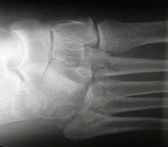
- Radiographs:
- fracture characteristics may be subtle;
- on non-stressed views, frx at base of 2nd metatarsal or anterior aspect of cuboid may most obvious
indications of Lisfranc injury;
- w/ questionable injury, consider wt bearing AP view to assess 1-2 interval;
- if standing AP is unacceptable to the patient then consider CT scan;
- intercuneiform region injuries: these may occur in upto 10-15 % of patients;
- lateral radiographs:
- lateral talometatarsal angle is formed by intersection of a line along the long axis of talus w/ long
axis of 1st metatarsal and normally forms a straight line
- ref: Prediction of midfoot instability in the subtle Lisfranc injury. Comparison of magnetic resonance imaging with intraoperative findings.
- Treatment of Sprains and Minimally Displaced Frx:
- Subtle injuries of the Lisfranc joint
- ref: Outcomes of Lisfranc Injuries in the National Football League
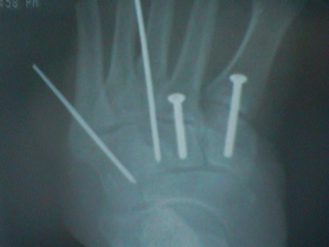
- Operative Treatment:
- Closed Reduction Percutaneous Pinning
- Open Reduction Internal Fixation:
- fractures presenting w/ more than than 2 mm of displacement and greater than 15 deg of
talometatarsal angulation require operative treatment;
- young competitive atheletes may require anatomic reduction;
- disrupted skin and excessive swelling are relative contra-indications for ORIF;
- note that pure dislocations w/o fracture may have a worse outcome despite ORIF;
- ref: Arthrodesis versus ORIF for Lisfranc fractures.
- Primary Arthrodesis:
- Salvage of Lisfranc's tarsometatarsal joint by arthrodesis.
- Severe Lisfrancs injuries: primary arthrodesis or ORIF?
- Open reduction internal fixation versus primary arthrodesis for lisfranc injuries: a prospective randomized study.
- Treatment of primarily ligamentous Lisfranc joint injuries: primary arthrodesis compared with open reduction and internal fixation. A prospective, randomized study.
- Arthrodesis versus ORIF for Lisfranc fractures.
- Does Open Reduction and Internal Fixation versus Primary Arthrodesis Improve Patient Outcomes for Lisfranc Trauma?
- post op:
- fixation must be rigid enough to prevent transverse plane & dorsoplantar motion of TMT joint and be maintained for at
least 12-16 weeks
Outcomes of Lisfranc Injuries in the National Football League.
Isolated fracture-dislocations of the first tarsometatarsal joint.
The diagnosis and treatment of injuries to the Lisfranc joint complex.
Fractures and fracture-dislocations of the tarsometatarsal joint
The treatment of tarsometatarsal injuries.
Fractures and fracture dislocations of the tarsometatarsal joint.
Functional outcome following anatomic restoration of tarsal-metatarsal fracture dislocation.