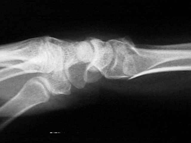
presents
Wheeless' Textbook of Orthopaedics
www.smith-nephew.com
Tracking Pixel
Search Site by Word
My Account

Lateral of C-spine


- See:
      - Flexion and Extension Views:
      - Technique:


- Upper Cervical Spine:
    - prevertebral soft tissues
    - occipital-atlanto-axial injury:
            - atlanto-occipital disassociation
                  - C1-C2 interspinous space should not be greater than 10 mm;
            - atlanto-axial impaction (rheumatoid C-spine)
            - atlas (Jefferson frx)
            - axis (odontoid frx / hangman frx)
            - atlantoaxial distance & SAC;
                  - ADI in children (< 10 yrs) < 3.5 mm; (see pediatric C-spine)
                  - ADI in adults < 3 mm;
                        - an anterior shift of C1 on C2 of more than 3-5 mm implies injury to
                                transverse ligament (see atlanto-axial subluxation);
                  - shift > 5 mm implies injury transverse & alar ligaments;
            - SAC:
                  - greater than 18   mm is normal normal;
                  - 15-17 mm - grey zone;
                  - less than 14   mm is consisent w/ cord compression;
    - pseudosubluxation of c spine:

- Sub-Axial Spine - Alignment:
    - posterior cortices: (more important than anterior cortices)
          - anterior or posterior translation of vertebral bodies > 3.5 mm implies instability;
          - w/ less than 25% relative shift of one vertebral body over another
                  consider facet frx;
          - w/ 25% relative shift consider unilateral facet dislocation
                  and w/ 50% shift, consider or bilateral facet dislocation;
    - vertebral body angulation / translation:
          - patterns of instability include:
                - 1.7 mm or greater of disk widening;
                - 3.5 mm of translational displacement;
                - angulation between two adjacent vertebra of 11 deg more than contiguous cervical vertebrae;
                - measurements are made from each inferior endplate;
    - anterior cortices:
          - anterior subluxation
          - minimal compression frx of anterior vertebral bodies;
          - tear drop sign: bone chip off antero-inferior aspect;
                - may indicate displacement of disc or posterior fragment of
                      vertebral body into spinal canal & cord injury;
    - spinolaminar line (dorsum of lateral masses) (see oblique view);
          - facet joint widening;
          - rotation of the facets on lateral view;
          - parallel articular process facets;
    - spinous process angulation:
          - C1-C2 interspinous space should not be greater than 10 mm;
          - widening is present when the distance is more than 1.5 times the
                inter-spinous distance of adjacent spinal segments;
          - fanning implies middle column disrupton;


Biomechanical analysis of clinical stability in the cervical spine.
    Clin Orthop Rel Res. 1975; 109: 85-96.
    White A., Johnson R., Panjabi M., and Southwick W.

Neurapraxia of the cervical spinal cord with transient quadriplegia.
    J Bone Joint Surg (Am) 1986;68A:1354-1370.
    Torg JS, Pavlov H, Genuario S, et al:


Original Text by Clifford R. Wheeless, III, MD.