

- Extensor Tendon Rupture
- Extensor Digitorum Communis
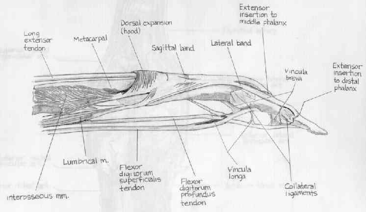
- Extensor Mechanism of Digits
- Anatomy of EDC:
- note that EDC is less tolerant of shortening than FDS due to differential in
tendon excursion (50 mm compared to 70 mm, respectively);
- references:
- The terminal tendon of the digital extensor mechanism: Part I, anatomic study.
- The terminal tendon of the digital extensor mechanism: Part II, kinematic study.
- A study of dynamic anatomy of extensor tendons and implications for treatment.
- Anatomy of the extensor tendons to the index finger.
- Zone Classification:
- zone I - mallet finger
- zone II - middle phalanx
- extensor apparatus extends over the dorsal half of the digit;
- at this point the lateral bands are traveling in a volar to dorsal direction, and are merging with fibers of the central slip;
- usually tendon injuries in this location are partial, since the extensor tendon extends over the dorsal half of the digit;
- at this level the lacerated tendon is repaired w/ 4-0 Vicryl figure of 8 sutures;
- zone III - PIP joint:
- zone IV: (proximal phalanx):
- at the level of the proximal phalanx, there is only 2-3 mm of tendon excursion, and therefore, minor amounts of adhesion can
result in significant amounts of loss of extension;
- usually the lateral bands are spared in this location due to their volar location, however it is important to repair them
separately if they are involved;
- tendon can be repaired w/ figure of eight 4-0 Ethibond sutures, but take care to avoid tendon shortening;
- PIP joint is held in extension for 6 weeks;
- zone V - over MP joint:
- exam for tendon injury can be confused by the action of the hand intrinsics (which have the effect of extending the digits);
- saggital band injury may also occur in Zone V injuries and require individual repair w/ interrupted-inverted 4-0 Ethibond sutures;
- failure to perform this repair may result in tendon subluxation;
- visualization is facilitated by longitudinally extending the wound;
- the tendon edges are retracted proximally and distally to expose the joint capsule;
- the capsular tear can be extended longituinally to allow joint washout;
- flexing the joint allows inspection of the metacarpal head;
- zone VI - dorsum of hand and wrist
- The Effect of Progressive Extensor Retinaculum Excision on Wrist Biomechanics and Bowstringing
- Considerations for Tendon Repair:
- partial lacerations:
- partial laceration of the flexor tendons or the extensor tendons proximal to the MP joint may or may not require repair;
- partial lacerations of the extensor tendons at or distal to MP joint level must be repaired;
- tendon repair techniques:
- note that the ultimate strength of a tendon repair depends on the number and size of sutures crossing the laceration site, where
as, resistance to gap formation depends on suture purchase;
- perhaps the easiest and quickest technique is a running horizontal mattress technique;
- this technique is especially well suited for tendon lacerations distal to the MCP joint, where the tendon is relatively flat;
- repair allows multiple suture strands to cross repair site, and when carefully performed, there is minimal tendon shortening;
- as noted by Newport and Williams (1992) Kleinert modification of the Bunnell technique is stronger than the modified Kessler
but both of these are significantly stronger than the horizontal matress and figure of 8;
- as noted in the report by Howard and Greenwald 1997, the MGH tendon repair technique (crossing running suture repair) was
signficantly more resistant to gap formation than the Bunnel or the Krackow technique;
- MGH tendon repair has superior suture purchase which is probably related to superior resistance to gap formation;
- Biomechanical characteristics of extensor tendon suture techniques.
- staged extensor tendon repair:
- consists of staged tendon reconstruction using a silicone implant;
- indicated for combined extensor injuries (skin, tendon, joint capsule, and bone ect);
- small skin incisions are made over the dorsum of the finger when necessary;
- silicone rod is placed along the pretendinous fascia to make a premade tunnel;
- the rod will help provide extension thru elastic recoil of the rod;
- soft tissue defects are managed by STSG or by secondary intention;
- once the soft tissues have healed, the silicone rod is exchanged for a tendon graft;
- references:
- Staged extensor tendon reconstruction in the finger.
- The use of silicone rods under split-thickness skin grafts for reconstruction of extensor tendon injuries.
- Post Operative Care:
- post operative rehab is dependent on the level of extensor tendon injury;
- in general, w/ extensor tendon injuries proximal to the MP joints rehab is dedicated to keeping the MP joints in extension for 1-2
weeks, followed by a passive extension/active flexion splint;
- PIP joints may remain free during the entire postoperative time period;
- in general, w/ extensor tendon injuries distal to the MP joints, the PIP and DIP joints are held immobilized in extension for about
4-6 weeks (as would be done for a mallet or boutoniere injury);
- references:
- Controlled mobilization of extensor tendon lacerations: Study of 120 cases. Allieu Y, et al. Rev Chir Ortho. 1984;70:68.
- Wrist position and extensor tendon amplitude following repair.
- Early dynamic splinting for extensor tendon injuries.
- Management and rehabilization of extensor tendon injuries.
- A comparison of results of extensor tendon repair followed by early controlled mobilisation versus static immobilisation.
- Dynamic traction after extensor tendon repair in zones 6, 7, and 8: a retrospective study.
- Results of dynamic splintage following extensor tendon repair.
- Complications:
- loss of flexion can occur due to extensor tendon shortening;
- loss of flexion and extension can result from adhesions;
- patient's may note a weakened grip;
- extrinsic tendon tightness:
- may result from crushing injuries to the hand;
- test extrinsic tightness by test PIP flexion while the MP joints are held flexed;
- w/ extrinsic tightness, there will be more PIP flexion w/ the MP's held extended;
- w/ the PIP joints fully flexed, the MP joints will move into extension;
- this requires aggressive hand therapy, and if no improvement is found, then extensor tendon tenolysis and dorsal joint
capsulotomy is required;
- in some cases a Littler Release may be appropriate:
- a portion extrinsic tendon is excised over the proximal phalax;
- extrinsic tendon will then extend the MP joint, and the intrinsic tendons will then extend the PIP joint;
- intrinsic tendon tightness:
- may result from crushing injuries to the hand;
- in contrast to extrinsic tendon tightness, when the MP joint is extended, flexion of the PIP joints is limited;
- extensor quadrigia effect:
- results from shortening of one tendon, and resultant loss of motion of the other extensor tendons (due to the interconnecting junctura)
Extensor tendon injuries.
Long-Term Results of Extensor Tendon Repair.
Biomechanical characteristics of extensor tendon suture techniques.
Extensor tenolysis: a modern version of an old approach.
Complications in phalangeal and metacarpal fracture management. Results of extensor tenolysis.
Biomechanical analysis of four-strand extensor tendon repair techniques.
Extensor tendon grafting on the dorsum of the hand in massive tendon loss.
Biomechanical analysis of four strand extensor tendon repair techniques.