

- Indirect Reduction Stratedgy:
- statedgy forgoes attempts at meniscal reduction (or repair), ligamentous repair, or direct joint reduction;
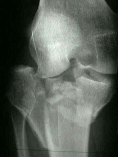
- longitudinal traction is used to align proximal tibial condyles over shaft;
- depressed fragments are reduced w/ a bone tamp thru cortical window;
- frx reduction is maintained w/ percutaneously appliede tenaculum clamp;
- note that flouroscopy may under-estimate joint incongruity as compared to postop plain radiographs;
- 6.5 cannulated screws are inserted percutaneously to hold reduction;
- these are placed immediately under the subchondral surface;
- EBI fixator (see: external fixators)
- applied anteromedially;
- two or three cancellous screws are placed in proximal tibia;
- pins should be placed at least 1 cm away from the joint surface but joint sepsis can occur w/ pins placed even 3 cm from joint
(due to joint capsule disruption);
- other option is to place pins in distal femur (anteromedially); which then requires a second operation for pin insertion into the
proximal tibia at a later date;
- three cortical screws are placed distally;
- post-op, it is essential to rule out proximal pin site infections which can lead to septic arthritis;
- dynamization of Ex-fix is carried out at 4-8 weeks