

- Exposure:
- similar to the ilioinguinal approach:
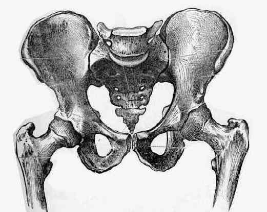
- it is necessary to expose the iliac crest & SI joint including the ala of sacrum;
- ensure that the patient is on a flouro table and that patient's pelvis is distal to the main table support (in order to get xrays);
- long incision is made from the anterior superior iliac spine to the posterior portion of the iliac crest and then up the flank;
- dissect down to the demarcation between the gluteus medius and abdominal muscles on the outer table of the iliac wing;
- incise through this avascular line, and then elevate the abdominal muscles and the iliopsoas off of the top of the crest and the inner table;
- iliac crest and its inner wall are exposed and the iliacus muscle swept by subperiosteal dissection posteriorly to expose SI joint, including ala of sacrum;
- inferiorly, the iliacus needs to be dissected down to the pelvic brim;
- more posteriorly, the dissection is essentially retroperitoneal, which allows the iliopsoas and the abdominal muscles to be retracted medially;
- this improves exposure of the sacral ala;
- greater sciatic notch is exposed to protect scaitic nerve;
- additional exposure is afforded by inserting one Homan retractor into the anterior sacral ala (medial to the SI joint) and a second homan retractor over the pelvic brim;
- hazards:
- L5 nerve root exits from the intervertebral foramen between L5 and S1 and crosses the L5-S1 disc to the ala of the sacrum, where it joins S1 nerve root as it exits from the S1 foramen;
- L5 nerve root lies about 2-3 cm from the SI joint;
- excessive medial retraction may injure this nerve route;
- note that the report by Atlihan, et al (2000), the authors report on 60 dissections which demonstrated that the L4 nerve root was most at risk during dissections over the anterior aspect of the anterior SI joint;
- ref: Anatomy of the anterior sacroiliac joint with reference to lumbosacral nerves.
- Reduction:
- secure SI joint anteriorly by approaching it from over iliac wing, which permits direct visualization of reduction & placement of short plates and screws;
- anatomical reduction with pointed reduction forceps in the anterior superior spine of the ilium pulling forwards;
- reduction is checked anteriorly at the greater sciatic notch;
- reduction care be held provisionally w/ K wires;
- alternatively, reduction is achieved by inserting a single screw in the ala and on in the ilium, and then compressing the screws w/ a reduction clamp;
- care not to damage any nerves, esp L5 root in front of SI joint;
- Surgical Fixation:
- apply a two or three hole 3.5 mm DCP, placed across anterior aspect of SI joint;
- uses two three-hole plates spanning from the anterior surface of sacral ala to internal iliac fossa, using 4.0 mm fully threaded cancellous bone screws;
- overcompression of the anterior aspect of the SI joint is common with plate fixation unless the plate contour is perfect;
- prior to screw insertion, identify the plane of the SI joint inorder to ensure that screws do not cross the joint;
- only one screw can be inserted into sacrum to avoid the L5 nerve root, and only one or two into the adjacent ilium since the bone rapidly become thin;
- alternatives:
- 7.0 mm cannulated screws or 6.5 mm cancellous bone screws as lag screws;
- Hazards:
- neurologic: care is taken to avoid stretching L-5 nerve root which passes across anterior aspect of the sacral ala
- vascular: Vascular risk reduction during anterior surgical approach sacroiliac joint plating
The Unstable Pelvic Fracture: Operative Treatment.