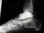
- Type I Talar Fractures:
- non displaced fractures of the talar neck without dislocation
- AVN: 10%
- important challenge w/ this injury is ensuring that anatomic reduction is obtained with no varus rotation;
- treated closed, usually w/ short leg cast w/ foot in slight equinus;
- leg is kept non-wt bearing for 4 wks, & then wt bearing is allowed in cast for another 8 wks or until healing is evident by x-ray (evidenced by trabeculations across fracture site);
- Type II Talar Fractures:
- displaced fracture of the talar neck with subluxation or dislocation of the sub-talar joint (subtalar joint subluxation is usually dorsal) and the ankle remains aligned;
- type II injuries are caused by the destruction of the talocalcaneal ligament;
- it is difficult to perform closed reduction of the dorsal and supination deformity;
- if closed reduction is possible then 8-12 weeks is necessary for healing (trabeculation across the fracture site);
- ORIF is needed if there is more than 3-5 mm of dorsal displacement and any rotational deformity;
- AVN: 30%
- Type III Talar Fractures:
- displaced frx of talar neck w/ dislocation of body of talus from both subtalar joint and the ankle joint;
- when body dislocates, it is usually found on posterior medial aspect adjacent to the Achilles tendon;
- in this location, there can be compression of neurovascular structures, & care must be taken when approaching by open means dislocated body of the talus;
- talocalcaneal ligament is ruptured when there is dorsal displacement of the distal fragment;
- after rupture of this ligament, it is difficult to control distal talar neck fracture by closed means;
- requires ORIF:
- retrograde K wires can be placed thru frx & out post-lateral aspect;
- cannulated screws can be used thru the posterior-lateral aspect using the wires;
- AVN: 90%
- Type IV:
- subtalar, tibiotalar, and talonavicular joint subluxation or dislocation (see subtalar dislocation);
- talar neck fracture w/ dislocation of the head fragment;
- open type IV fractures are associated w/ high rate of infection (30%), despite aggressive debridement and infection;
- salvage treatment:
- consider placement of methylmethacrylate spacer shaped like a talus;
- there are documented cases of patients being pain free for several years with this method of treatment;
- Hawkins Sign:
- appearance of decreased subchondral bone density in the dome of talus 6 to 8 weeks following injury indicates that there is sufficient
vascular supply to bone to allow normal disuse osteopenia to occur;
- resorption of subchondral bone is a consequence of disuse osteoporosis and suggests that the bone segment has adequate circulation, and that normal healing is occuring
Fractures of the neck of the talus.

