

- Determine Congruency
- wt bearing AP & lateral radiographs:
- IMA: (nl < 10 deg)
- note that this angle is highly dependent on the technique of measurement
- some authors measure from down the metatarsal shaft, where as, other authors measure from the center of the metatarsal base to the center of the metatarsal head;
- distal metatarsal articular angle:
- normally this is zero deg;
- lateral deviation more than 10 deg is abnormal;
- typically a moderately severe hallux valgus w/ a significantly increased DMA will be associated w/ a congruent bunion;
- hallux valgus angle:
- normal < 15-18 deg;
- when the valgus angle of the 1st MTP joint > 30-35 deg, pronation of the great toe results and other structures are also affected (plantar shift of abductor hallucis & lateral shift of sesamoid & intrinsics, and often hammering of the second toe);
- first metatarsocuneiform joint angle:
- high angle of inclination or presence of a lateral facet at the base of the 1st MT shaft;
- this mechanically blocks the the metatarsal from being brought into satisfactory alignment unless an osteotomy of the first metatarsal is carried out;
- sesamoids:
- moderate subluxation:
- lateral sesamoid is uncovered 50 to 75 % within 1st IM space;
- medial sesamoid is located in a central position plantar to the first metatarsal head;
- severe subluxation:
- lateral sesamoid moves to the lateral aspect of the first metatarsal head is dorsal to the medial sesamoid;
- Misc:
- generalized metatarsus adductus:
- lengths of the 1st & 2nd metatarsals:
- 1st talometatarsal angle (lat. x-ray):
- degree of hallux interphalangeus
- size of Medial Eminence
- evidence of arthrosis of the 1st MP joint
- obliquity of the first MT-cuneiform joint
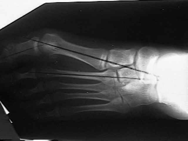
Radiographic analysis of hallux valgus. A two-dimensional coordinate system.