lateral incision over distal fibula, which is extended anteriorly at distal tip of fibula toward the base of 4th metatarsal;
be aware of peroneal tendons and note that the sural nerve lies 2 cm below tip of fibula;
dissect down to the fibula and elevate the periosteoum both anteriorly and posteriorly;
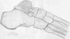
expose the sinus tarsi and the posterior facet of the subtalar joint;
remove contents of sinus tarsi;
anteriorly, expose the lateral and anterior aspect of the tibia and ankle joint;
identify talar neck, elevate the extensor tendons, and insert small spade retractor to protect N/V bundle;
management of the fibula:
osteotomy and retention of the fibula (fibula provides lateral support of the fusion);
fibula is transected well above the ankle joint, and the inner (medial) half is removed with a saw;
fibula is opposed to the tibia and talus with 3.5 screws (syndesmosis is fused);
care is taken to avoid posterior soft tissue envelope dissection;
removal of the fibula
alternative procedure
consider transecting the fibula in an oblique: proximal-lateral to distal-medial direction;
subluxation of the peroneal tendons does not appear to be a problem;
Medial Incision: (used only if external fixator is not used);
make anteromedial incision over medial malleolus similar to the incision used for medial malleolar frx;
be aware of the saphenous vein anteriorly and the tibialis posterior which lies posteriorly;
excise periosteum and deltoid ligament;
Homan retractors are placed around the medial malleolus;
transect the medial malleolus at the level of the plafond;
removing the medial malleolus will narrow the ankle which will facilitate shoe wear, and in addition resection of the medial malleolus leads to better tibio-talar opposition;
the anterior 2/3 of the medial malleolus is expendable in ankle fusions;
the posterior 1/3 may serve to protect the tendons and N/V bundle;
cut at oblique 45 deg angle from a line parallel w/ the tibia;
note that Mann and Rongstad 1998 determined that greater resection of the medial malleolus correlated to a longer healing time and to an increased rate of nonunion;
these authors also emphasize preservation of the deltoid ligament, inorder to preserve the blood supply to the talus;
reference:
Arthrodesis of the Ankle: A critical analysis. RA Mann MD and KM Rongstad MD. Foot and Ankle International. Vol 19. No 1. Jan 1998. p 3-9.
Exposure of Ankle Joint:
free up ankle attachments;
either sublux or dislocate ankle joint in a medial direction;
joint is exposed thru the lateral wound;
Removal of Cartilage from Ankle Joint:
remove the cartilage and subchondral bone from the distal tibia;
shortening of extremity occurs with more bone resection;
some argue, however, that because the subchondral bone at the distal tibia is especially thick, that the distal cut should be made upto the point of the central concave surface of the distal tibia;
in some cases, a high speed burr is useful to remove bone from the posterior tibial plafond;
Talar Cut;
before resecting talar cartilage, consider pinning talus to transected surface of distal tibia, to determine optimum position;
once, optimum position is achieved, begin cut into talus at a depth of 3mm w/ blade directed parallel to distal tibial surface;
then retract pin and complete cut;
all articular cartilage and subchondral bone must be removed;
talus is relatively avascular, and bleeding cancellous bone w/ tourniquet in place may not be present;
Pitfalls:
avoid over-resection of the subchondral joint surfaces inorder to preserve ankle joint height as much as possible;