

- protocols: fat suppressed T2 wt images and proton density weighted images
- older protocol: T1 & T2 coronal oblique spin-echo sequences with use of 14 cm field of view and a four-millimeter slice thickness;
- Discussion:
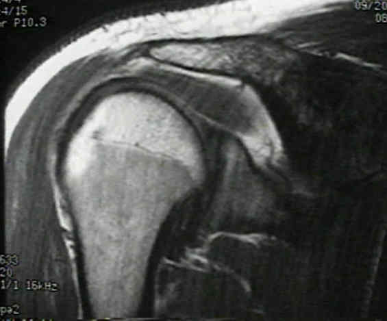
- it is the most important view for identification of a lesion of rotator cuff, as supraspinatus and infraspinatus are seen in continuity w/ their insertion on greater tuberosity;
- axis is parallel to supraspinatus muscle and tendon;
- infraspinatus is seen on posterior images and is more obliquely oriented;
- it shows relationship between supraspinatus & acromion (& AC joint);
- sagittal oblique plane, allows optimum visualization of supraspinatus outlet and of the shape of the acromion;
- coracoclavicular, coracohumeral, & CA ligaments can sometimes be seen;

- Anterior Coronal Oblique Images:
- supraspinatus tendon may curves anteriorly, which may cause averaging of images w/ rotator interval which may cause an increased signal and false impression of rotator cuff tear

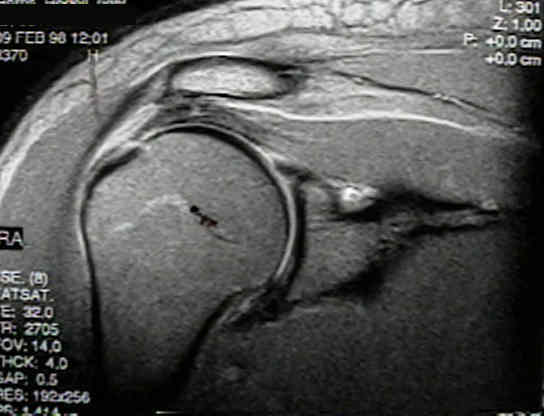
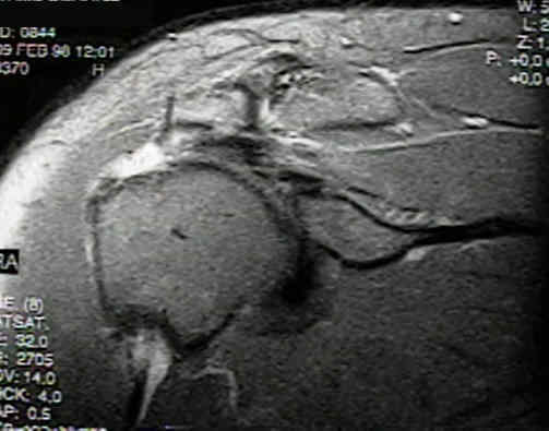
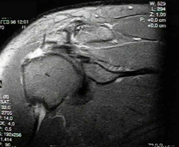
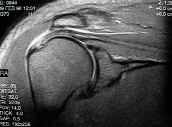
- Case Example: (55-year-old female with large rotator cuff tear which lead to anterior instability)
Abnormal findings on magnetic resonance images of asymptomatic shoulders.
Magnetic resonance imaging of the shoulder. Sensitivity, specificity, and predictive value.
Magnetic resonance imaging of the shoulder.
Shoulder instability: evaluation with MR imaging.
The use of MRI about the shoulder. Beltran J. J Shoulder Elbow Surg. 1992;1:321.