

- History:
-Maurice Klippel and Andre Feil first to describe syndrome in 1912
-Characterized by patients with:
Short neck
Low hairline
Decreased cervical motion
-Fewer than 50% with congenital defects of cervical spine have all three signs
- Discussion:
- involves congenital failure of segmentation of cervical vertebrae;
- results from failure of normal segmentation of cervical somites at 3-8 weeks's gestation;
- result is multiple fused cervical segments;
- spectrum of deformity from fusion of 2 vertebrae to involvement of entire C- spine;
- fusion of C-2 & C-3 is most common;
- familial Klippel-Feil-syndrome gene locus on the long arm of chromosome 8;
- associated conditions:
- consistently associated with congenital anomalies of other systems;
- congenital scoliosis;
- seen in 60%;
- majority require treatment;
- Sprengel's deformity; (33%)
Failure of scapula decent
Attached to cervical spine by omovertebral bone or fibrous band
- renal dz:
- occurs in 33%
- aplasia is common;
- renal ultrasound in indicated;
- synkinesis (mirror motions);
- congenital heart dz;
- brain stem abnormalities;
- congenital cervical stenosis;
- syndactyly and hypoplastic thumb;
- hearing loss: may occur in 30% of patients;
- ref: Klippel-Feil syndrome and deafness. A study with polytomography.
- diff dx:
- juvenile rheumatoid arthritis;
- rheumatoid spondylitis;
- Clinical Presentation:
-Often incidental finding
-Anomalies present at birth
-Diagnosed at later age
-Presentation:
Abnormal head position
Torticollis
Restricted Cervical ROM
Patients with extensive fusions present earlier
Cosmetic deformity
Instability/Hypermobility at unfused levels
Cord compression in congenitally anomolous, narrow canal in young adults
-Arnold Chiari Malformation
Ataxia
Dizziness
Nystagmus
-CNS involvement
Difficulty swallowing
Disturbed phonation
Hydrocephalus
Blurred vision
Headache
-Vertebral artery involvement
Rare
Syncope, Seizures, Ataxia
- Clinical Findings:
- low posterior hairline;
- short neck;
- limited neck range of motion (esp lateral side bending);
- scoliosis:
60% with curve >15°
Congenital or compensatory
Winter, et al. (1984) reported 25% incidence of KFS in 1215 patients with congenital scoliosis
- Classification:
Type I: Massive fusion of cervical spine
Type II: Fusion of one or two cervical interspaces
Type III: Thoracic or Lumbar vertebrae involved
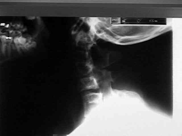
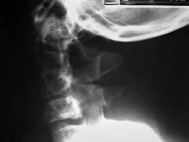
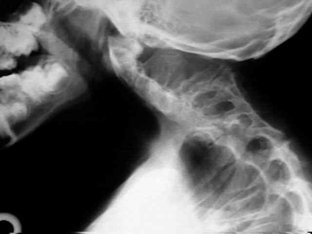
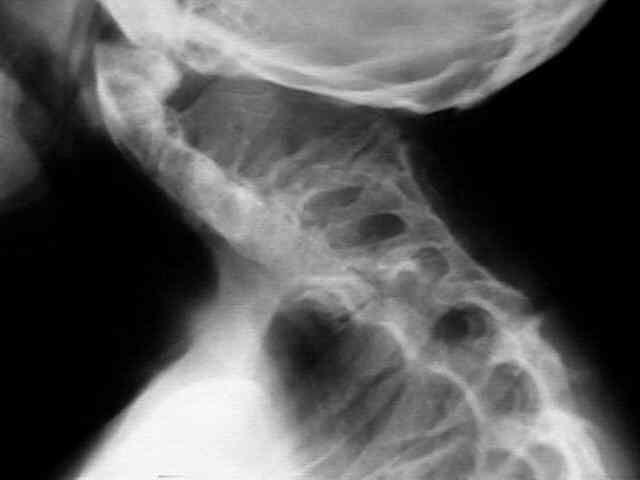
- Evaluation:
-C-spine series with flexion and extension
-Location & number of fused segments
-75% occur in C1-C3; C2/3 most common
-50% involve ≤ 3 Vertebrae
-Interspinous distance on flex-ex
-Translational instability
-Hemivertebrae/block vertebrae
-T&L spine imaging
-CT scan
Valuable tool to assess the bony anatomy
Helpful in pre-operative planning
Obtain reconstructions
-MRI
Identifies spinal cord abnormalities
Flexion and extension MRI to identify cord compression or stenosis
Ritterbusch et al.
25% with 5 mm or more of C1-C2 subluxation
25% with stenosis/12% had cord abnormalities
- Natural History: Pizzutillo, et al. (1994)
-Evaluated flex-ext radiographs to determine
-Alterations from normal motion
-Potential neurologic risk
Conclusion:
If hypermobility of upper cervical segment, greatest risk of neurologic sequelae.
If hypermobility of lower cervical segment, greatest risk of degenerative changes.
- Treatment:
If asymptomatic & no evidence of instability:
-Periodic flex-ex radiographs
-Avoidance of contact sports
-Avoidance of occupations and recreational activities with risk of head trauma
Sports Participation:
Type I
Absolute contraindication to participation in contact sports
Type II
Absolute contraindication
Fusion of one or two interspaces with:
-Associated limited motion
-Occipitocervical anomalies
-Involvement of C2
-Instability
-Disc disease
-Degenerative changes
No contraindication
Fusion of one or two interspaces at C3 or below with:
-Full cervical ROM
- Absence of above
Symptomatic Treatment:
-Modification of activities
-Bracing
-Traction
-Reduce symptoms
-Delay surgery
-Prevent neurologic compromise after minor trauma
Surgical intervention considered for:
-Progressive symptomatic segmental instability
-Neurologic compromise
Techniques:
-Occipitocervical arthrodesis
-Halo immobilization
-Atlantoaxial arthrodesis
Gallie
Brooks
Mageryl
-Subaxial
Interspinous
Sublaminar
The incidence of Klippel-Feil syndrome in patients with congenital scoliosis and kyphosis.
Risk factors in Klippel-Feil syndrome.
Klippel-Feil syndrome. A constellation of the associated anomalies.
Klippel-Feil Syndrome: Clinical Features and Current Understanding of Etiology.