

- See: Extensor Pollicis Longus Rupture:
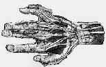
- Anatomy:
- origin: posterior surface of the middle 1/3 of the ulna & interosseous membrane;
- insertion: posterior surface of the base of the distal phalanx of thumb;
- action: extends the distal phalanx of the thumb; continued action, extends proximal phalanx and adducts the 1st metacarpal;
- nerve: PIN branch of deep radial nerve, C6, C7, & C8;
- Tunnel III:
- on ulnar side of Lister's Tubercle contains EPL , which defines ulnar border of anatomic snuff box;
- EPL tendon takes 45 deg turn around Lister's tubercle;
- then after passing over ECRL & ECRB tendons of tunnel I, it continues along its course to the thumb;
- note: that the "cross over" between the EPL and the ECR tendons can become involved in a cross over syndrome, just distal to the
extensor retinaculum;
- Exam:
- palpate length of tendon, look for any signs of rupture;
- ask pt to place hand flat on table, & lift only thumb off surface;
- w/ rupture, patient will be unable to raise the thumb in line w/ the second metacarpal