

Discussion
- tears of AC & CC ligaments (from fall on tip of shoulder) allows upper limb to drop away from clavicle, producing separation of AC joint;
differential diagnosis
- distal clavicular physeal separation
- childhood equivalent of AC separation;
- atraumatic AC joint laxity (from ligamentous laxity)
Classification
Rockwood Classification
- type I
- sprain of joint with out a complete tear of either ligament
- type II
- tear of AC ligaments w/ coracoclavicular ligaments intact;
- will not show marked elevation of lateral end of clavicle;
- type III
- in this injury both AC & CC ligaments are torn;
- > 5 mm elevation of AC joint w/o weights is consistent w/ severe type II or a type III injury;
- need to distinguish this from type III clavicular fracture
- type IV
- distal clavicle impaled posteriorly into trapezial fascia;
- type V

Basamania Classification
- essentially relies on whether the distal clavicle is stable or unstable;
- w/ more than 50-75 % displacement on static films or more than 100% displacement on a cross arm AP, there will be disruption of not only the AC ligaments but also the CC ligaments;
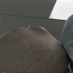
- clinically, an unstable AC separation will cause significant prominence of the distal end of the clavicle when the arm is distracted in adduction;
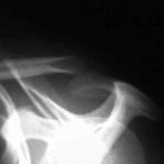
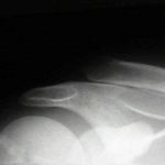
Radiograph
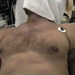
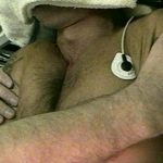
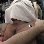
- Cross Body Adduction View
- (from C.J. Basamania MD personal communication, 1997);
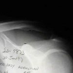
case example
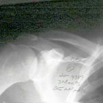
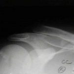
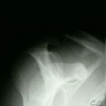
- 20-year-old who fell on tip of right shoulder, but did not show radiographic signs of AC joint injury in the ER;
- one month later the patient continued to have pain, and radiographs demonstrated greater than 100 percent displacement of AC joint on both AP and Cross Body AP (Cross Adduction View);
Operative Treatment
References
- Surgical treatment of acute type-V acromioclavicular injuries in athletes.
- Four-year outcome of operative treatment of acute acromioclavicular dislocation.
- Acromioclavicular joint injuries.
- A classification of acute acromioclavicular dislocation: a clinical, radiological and anatomical study.
- Late reconstruction of the ligaments following acromioclavicular separation.
- Acute, complete acromioclavicular separation.
- Conservative treatment of grade III acromioclavicular dislocations.
- Percutaneous cannulated screw coracoclavicular fixation for acute acromioclavicular dislocations.
- Complete acromioclavicular separations. A comparison of operative methods.
- Dislocation of the acromioclavicular joint. An end-result study.
- Conservative or surgical treatment of acromioclavicular dislocation. A prospective, controlled, randomized study.
- Year Book: Four-Year Outcome of Operative Treatment of Acute Acromioclavicular Dislocation.
- Acute dislocation of the acromioclavicular joint. Traumatic anatomy and the importance of deltoid and trapezius.
- Repair of complete acromioclavicular separations using the acromioclavicular-hook plate.
- Surgical treatment of acute type-V acromioclavicular injuries in athletes.
- Comprehensive functional analysis of shoulders following complete acromioclavicular separation.
- Conservative or surgical treatment of acromioclavicular dislocation. A prospective, controlled, randomized study.
- Surgical treatment of acute type-V acromioclavicular injuries in athletes.
- Radiological evaluation of the acromioclavicular joint.
- Biomechanical study of the ligamentous system of the acromioclavicular joint.