

- See:
- Pediatric Distal Tibial Fracture
- Pronation-External Rotation Frx
- Supination - External Rotation Frx
- Supination Inversion Frx
- Supination Plantar Flexion
- Tillaux Fracture
- Triplane Fracture
- Discussion:
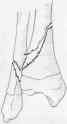
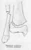
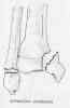
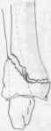
- most common epiphyseal injury to ankle is SH type II of distal tibia, caused by supination and external rotation;
- deltoid and lateral ligaments of ankle joint insert into epiphysis of distal tibia and fibula, and because, ligaments are stronger than the phsysis,
which accounts for the high occurance of SH type III and IV frx;
- pediatric ankle frx are most common between 10-15 years of age, as the growth plates begin to fuse;
- over a 1 1/2 year period, the tibial growth plate first closes in the mid region, then closes medially, and finally closes laterally;
- references:
- Classification of ankle fractures in children.
- Physeal injuries of the ankle in children: classification.
- Children's ankle fractures. Classification and epidemiology.
- Clickable Map:
- Radiographs:
- oblique radiographs help distinguish SH type III from type IV frx;
- references:
- Growth plate fractures of the distal tibia: is CT imaging necessary?
- Prevalence and clinical significance of occult fractures in children with radiograph-negative acute ankle injury. A meta-analysis.
- Non Operative Treatment: (SH I and SH II frx)
- SER, PEER, and SPF injuries (w/ Salter Harris type I or II components) are usually treated by closed reduction & cast immobilization;
- inadequate reduction may be due to interposed periosteum, which then requires open and anatomic reduction;
- note: unlike SH III and SH IV frx, there is no indication that anatomic open reduction will decrease incidence of growth arrest in SH II ankle frx;
- Closed Reduction:
- closed reduction is usually adequate in all Salter Harris type I & II fractures of the distal tibial and fibular epiphysis;
- growth arrest may occasionally occur even in these frxs, since significant compression may have occurred at the time of injury;
- in young pt, < 10 yrs old, some displacement may be accepted since it will remodel with further growth;
- this is esp true w/ pronation-external rotation frxs which shows gradual correction of valgus deformity over 6 to 12 months;
- reduction must be performed gently, to avoid growth plate damage;
- reduction may be done under GEA;
- LLC are used in rotational injuries (SER, PEER) / SLC is used for SPF;
- at 3 wks cast is exchange for SLC (used for 3 more wks)
- Operative Treatment: (SH III and IV frx)
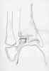
- Salter Harris III and IV fractures (Sup Inv Frx) require anatomic reduction (ref);
- may be accomplished w/ closed or, more often, w/ open reduction;
- use medial incision, centered over frx;
- avoid damage to perichondrium around the physis;
- either use 2 smooth K wires or cancellous screws;
- pins are directed from medial malleolus to tibial metaphysis, parallel to both the joint surface and the physis;
- note that the distal tibial articular surface is dome-shaped making it possible for pins to encroach into the joint;
- lateral radiographs help avoid this complication;
- cancellous screws are removed at 3 to 6 months after frx has healed;
- postoperatively, a LLC is used, which may need to be changed at end of first weeks due to swelling;
- references:
- Distal tibial physeal fractures in children that may require open reduction.
- Operative treatment of ankle fractures in children.
- Salter-Harris Type-IV injuries of the distal tibial epiphyseal growth plate, with emphasis on those involving the medial malleolus.
- Intraoperative arthrography for evaluation of closed reduction and percutaneous fixation of displaced MacFarland fractures: an alternative to open surgery.
- Complications: Growth Plate Arrest:
- cessation of growth plate does not occur immediately after injury to physeal plate;
- growth arrest may occur even in seemingly benign frxs, since significant compression forces may have been sustained at the time of injury;
- growth arrest may be delayed for 6 months or longer;
- hence, ankle frxs should be followed for approx 2 yrs;
- look for Harris's growth arrest line to confirm impending growth arrest;
- SH I and II frx: growth plate arrest is usually central;
- ref: Salter-Harris I and II fractures of the distal tibia: does mechanism of injury relate to premature physeal closure?
- SH III and IV frx: growth plate arrest is usually peripheral and more malignant;
- references:
- Salter-Harris Type-IV injuries of the distal tibial epiphyseal growth plate, with emphasis on those involving the medial malleolus.
- Premature physeal closure following distal tibia physeal fractures: a new radiographic predictor.
- Angular Deformities: due to asymmetric arrest of distal tibial growth plate:
- varus deformity;
- common angular deformity, frequently seen after supination inversion frx;
- an osseous bridge in the medial part of the physeal plate can develop after a Salter Harris III or IV injury;
- even initially undisplaced frx can develop a varus deformity;
- consider immediate growth arrest once varus deformity is noted;
- valgus Deformity:
- 2nd to asymmetric arrest of distal fibular growth plate:
- Rate of Correction and Recurrence of Ankle Valgus in Children Using a Transphyseal Medial Malleolar Screw
- malunion (pronation-external rotation Frx)
- references:
- Acquired valgus deformity of the tibia in children.
- Changes in tibiofibular relationships due to growth disturbances after ankle fractures in children.
- Prediction of growth pattern after ankle fractures in children.
- Leg Length Discrepancy: resulting from ankle frx;
- seen in about 10-30% of cases;
- avg discrepancy is only 1 cm, since distal tibial physis grows 2-3 mm/year;
- if the expected discrepancy is > 2 cm, an epiphysis of promixal tibial and fibula at the appropriate age is indicated;
- Avascular Necrosis
- Nonunion
Secondary Ossification Centers in the Development of the Medial Malleolus
Traction apophysitis of the medial malleolus.
Plantar flexion injuries of the ankle. An experimental study.
Fractures of the distal tibial epiphysis in adolescence.
Late results in 65 physeal ankle fractures.
Avulsion fracture of the lateral malleolus in children.
Fracture of the lateral portion of the distal tibial epiphysis.
Outcome of Physeal and Epiphyseal Injuries of the Distal Tibia with Intra-Articular Involvement.
Distal tibial physeal fractures in children that may require open reduction.
Fractures of the Distal Tibial Metaphysis in Children: Patterns of Injury and Results of Treatment.
Surgical Indications for Distal Tibial Epiphyseal Fractures in Children