

- Anterior Talar Impingement Syndrome
- Avascular Necrosis and Salvage of Talus Fractures
- Blood Supply to Talus
- Congenital Vertical Talus
- Talar Head Frx
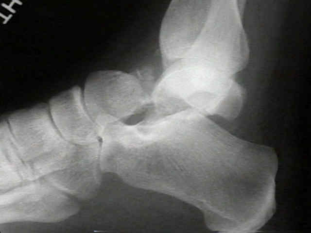
- Fractures of the Talar Neck:
- Hawkins Classification:
- Type I Talar Fractures
- Type II Talar Fractures
- Type III Talar Fractures
- Hawkins Sign
- Lateral Talar Process Frx
- Posterior Process Talus Frx
- Osteochondral Lesions of the Talus
- Radiographic Evaluation
- Sub Talar Dislocation
- References
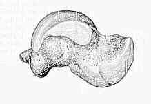
- Anatomy: 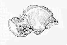
- talus is the highest portion of the dome, forms the joint w/ tibia and fibula, is supported by the other tarsal bones, and articulates w/
calcaneus below and navicular in front;
- presence of a secondary center of ossification in the lower end of femur and the upper end of tibia at about the time of birth, &
appearance of primary centers of ossification in calcaneus and talus in the sixth to the eighth fetal month (or earlier), and in
cuboid at or soon after birth, are data useful in medicolegal decision as to whether a newborn child is a full-term fetus;
- talus has a curved head, an intermediate neck portion and a large trapezoidal body;
- it articulates with the navicular, calcaneus, tibia, and fibula;
- body of the talus is almost entirely covered by articular cartilage;
- superior surface is convex from front to back, and slightly concave from side to side;
- dome of talus is trapezoidal in shape, and its anterior surface is an average of 2.5 mm (range 0-6 mm) wider than posterior surface;
- articular saurfaces of malleoli are also wider anteriorly and support the talus;
- medial & lateral articular facets of talus are continuous w/ superior articlar surface, & lateral facet is larger than corresponding facet
on the fibula;
- majority of the talar neck has no articular surface and serves as site of access for much of the blood supply to the rest of the talus;
- multiple articular facets and lack of muscular attachments are evidence of intercalary role of the talus in connecting the leg to foot;
- talocalcaneal ligament is important in the dorsal and supination displacement of a talar neck fracture;
- disruption of talocalcaneal ligament allows displacement of the frx;
- when the ligament is intact, it is possible to do a closed reduction;
- Mechanics of Talus:
- transverse tarsal joint (talus-navicular, calcaneal-cuboid)
- motion is based on foot position w/ 2 axises of rotation (talo- navicular and calcaneocuboid)
- w/ eversion of foot (as in early stance phase), two joints are parallel and ROM is permitted;
- w/ foot in inversion (late stance), external rotation causes the joints to no longer be parallel, & limited motion is allowed;
- neck of talus, which is constricted inferiorly, laterally, & superiorly, is roughened by ligamentous attachments and
vascular foramina;
- it deviates medially approximately 15 deg to 20 deg in the adult and is the area of bone most vulnarable to fracture