

- See:
- Growth Deformities of Limbs
- Growth Plate Arrest
- Discussion:
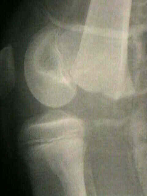
- anatomy of distal femoral physis:
- account for approx 5% of all physeal frx;
- displacement in sagittal plane is assoc w/ N/V injury in popliteal fossa and instability on closed reduction;
- common mechanism is hyperextsion causing anterior displacement of epiphysis;
- displacement in coronal plane is not assoc w/ other injuries, & joint may be stable after closed reduction.
- unlike most Salter-Harris type I physeal fractures, distal femoral physeal fracture course through multiple layers of the growth plate;
- additional stability occurs at this growth plate because of undulating topography and presence of mammillary bodies (prominences that interdigitate with other portions of the physis);
- Physical Exam Findings:
- pts usually are unable to walk or bear wt on injured extremity;
- hamstring spasm causes knee to be held in flexion;
- thigh may appear angulated & short compared w/ contralat thigh;
- pain, knee effusion, & soft-tissue swelling usually are severe;
- hemarthrosis may be more severe in SH III and IV fractures;
- anterior dimpling may be seen w/ hyperextension injuries;
- vascular exam may reveal diminished or absent pulses;
- w/ diminished pulses consider femoral arteriogram (arterial injuries associated w/ fractures);
- hyperextension deformity is more commonly associated with vascular injury;
- sensation on dorsal & plantar aspects of foot may be abnormal if posterior tibial or peroneal nerve has been damaged;
- Non Operative Treatment:
- non-displaced fx are treated w/ immobilization in an LLC or Single Leg Hip Spica for 4-6 wks
- be aware that displaced fractures may tend to redisplace without some form of internal fixation;
- acceptable reduction:
- posterior angulation upto 20 deg will remodel in kids < 10 yrs old,
- adolescent, however, will not remodle and will not tolerate this degree of angulation;
- no > 5 deg of varus-valgus angulation is acceptable;
- Salter Harris Type II Fractures:
- displaced SH type-I or II frx are reduced closed w/ pt under GEA;
- SH III and IV:
- tense hemarthrosis may require preoperative aspiration;
- require anatomic reduction, which can not be obtained w/ close reduction;
- even minimally unstable fractures can be unstable;
- Operative Treatment of Salter Harris Type II Fractures
- Operative Treatment: SH III and IV frx:
- operative treatment is required since, even slight physeal displacement can result in formation of osseous bar that causes limb-length discrepancy & angular deformity;
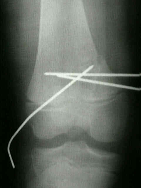
- if minimally displaced, SH III and IV frx are treated by percutaneously manipulating fragment w/ pin;
- following reduction, pin is driven across frx, parallel to to physis;
- most often open reduction and fixation is required for an anatomic reduction;
- a large (2-3 cm-high) triangular metaphyseal spike may be amenable to fixation w/ 2 cannulated 4.0 or 6.5-mm screws, inserted transversely to fix spike to metaphysis of femur w/o crossing physis.
- in other cases, pins or cannulated screws are inserted between the joint and the physis;
- long leg cast is applied with the knee in 5-10 deg of flexion.
- if fixation is not used, then immobilize in spica cast, w/ frequent f/u x-rays to detect any slippage or loss of reduction;
- references:
- Salter-Harris type III fractures of the distal femur: plain radiographs can be deceptive
- Salter-Harris type-III fracture of the medial femoral condyle occurring in the adolescent athlete.
- Complications: Growth Plate Arrest:
- limb length descrepancy of more than 1 cm may occur in over 40%.
- angular deformities may occur in a third of patients
Injuries of the distal femoral growth plate and epiphysis: should open reduction be performed.
Periarticular fractures after manipulation for knee contractures in children.
Traumatic injuries of the distal femoral physis. Retrospective study on 151 cases.
Predicting the Outcome of Physeal Fractures of the Distal Femur.
The effect of percutaneous pin fixation in the treatment of distal femoral physeal fractures