


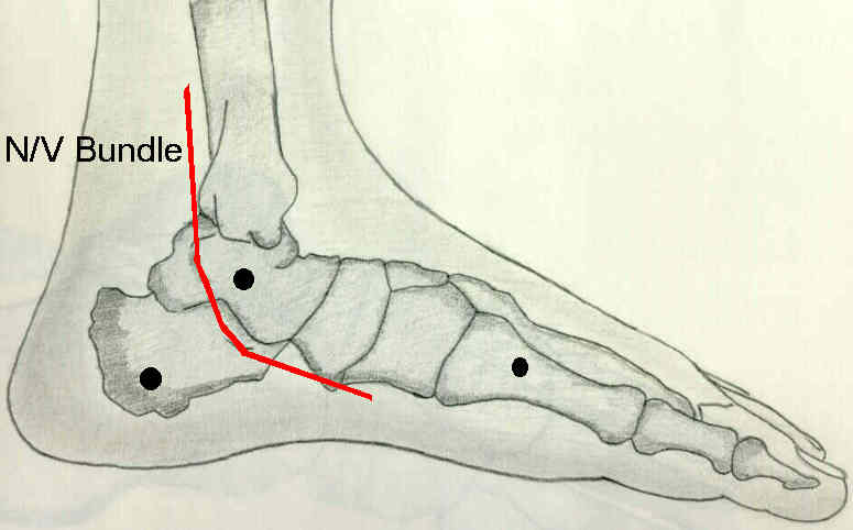
- General Considerations:
- safe zones for pin insertion imply that pins will avoid major neurovascular structures;
- besides avoiding neurovascular strucutures, it is important that pins be inserted in subcutaneous locations;
- subcutaneous insertion avoids muscular impalment which decreases pain and increases function;
- other advantageous include decreased swelling and decreased pin infection;
- Proximal to the Tibial Tubercle:
- pins can safely be inserted within an arc of 220 deg;
- safe zone extends from posteromedial border of tibial plateau to the tibiofibular joint (of course, this excludes the patellar tendon);
- transfixation wires:
- can be inserted through the anterior portion of fibular head, aiming 30 deg anterior across proximal tibia (from lateral to medial), to exit medial to the patellar tendon;
- a second wire is inserted transversely, anterior to MCL and head of fibula;
- halfpins:
- are inserted at the oblique lateral or medial aspect of proximal tibia;
- transfibular half pin:
- provides additional fixator stability;
- insert a guide wire through the fibular head and is driven out of the proximal tibia;
- insert a cannulated drill over the guide wire (drilling from the tibia into the fibula);
- the drill is removed and an appropriately sized half pin is inserted thru the tibia into the fibular head;
- hazards:
- all neurovascular structures are located posterior to fibular head and the MCL;
- because the synovial recess extends below the joint line, wires should be placed at least 1 cm below joint line to avoid potential septic arthritis;
- further, w/ tibial plateau frx, disruption of the joint capsule will mean that even pins placed 2-3 cm from the joint, will be ask risk for septic arthritis;
- you only need to see one of these to believe it;
- Just Distal to the Tibial Tubercle: 
- safe arc of insertion is decreased to 140 deg, and this level is not entirely safe for transfixion wires;
- anterior tibial artery is located just anterior to interosseous membrane, and the posterior tibial artery lies behind the tibialis posterior;
- therefore, transfibular transfixation pins cannot be inserted at this level;
- posteriorly directed pins are to be especially avoided;
- halfpins:
- are inserted at the oblique medial aspect of tibia;
- transfixation wires:
- are inserted at the oblique lateral aspect of tiba just distal to the tibial tubercle, (thru tibialis anterior) and exits along the postero-medial aspect of tibia;
- take care not to injure the saphenous vein or nerve;
- Mid-Tibia and Distal Tibia:
- along the distal tibia, the anterior tibial vessels hug the medial tibial cortex;
- transfixation wire is inserted 1 cm lateral to the tibial crest and is aimed posteromedially at an angle of 40-50 deg so that it passes just anterior to the posterior compartment musculature;
- generally this wire will pass posterior to the saphenous vein and nerve;
- if needed, a second wire can be inserted transversely from lateral to medial, thru the anterior compartment musculature and out the medial side of the tibia;
- Distal Tibia (above the ankle): 
- the safe arc remains at 120 to 140 deg, but the anterior tibial vessels and deep peroneal nerves become vulnerable as they cross the lateral tibial cortex;
- transfixation wires:
- transfibular fixation wire: placed thru anterior portion of fibula (to avoid the peroneal vessels);
- transverse fixation wire: avoid transfixation of the saphenous nerve and vein;
- may be inserted from lateral to medial, starting at a point 1 cm anterior to the fibula;
- alteratively insert a wire from lateral to medial starting at a point just lateral to the tibialis anterior (note that the vessels will lie just lateral to this point) and aiming posteromedially to exit the tibia just in front of the tibialis posterior;
- transfibular half pin:
- provides additional fixator stability;
- insert a guide wire through the fibula and is driven out of the proximal tibia;
- insert a cannulated drill over the guide wire (drilling from the tibia into the fibula);
- the drill is removed and an appropriately sized half pin is inserted through the tibia into the fibula;
External fixation of the tibia. Basic concepts and prospective evaluation.
Soft tissue injuries with the use of safe corridors for transfixion wire placement during external fixation of distal tibia fractures: an anatomic study.
Evaluation of Popliteal Artery Injury Risk With Locked Lateral Plating of the Tibial Plateau