

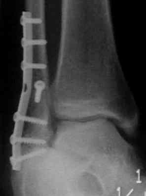
- Radiographs:
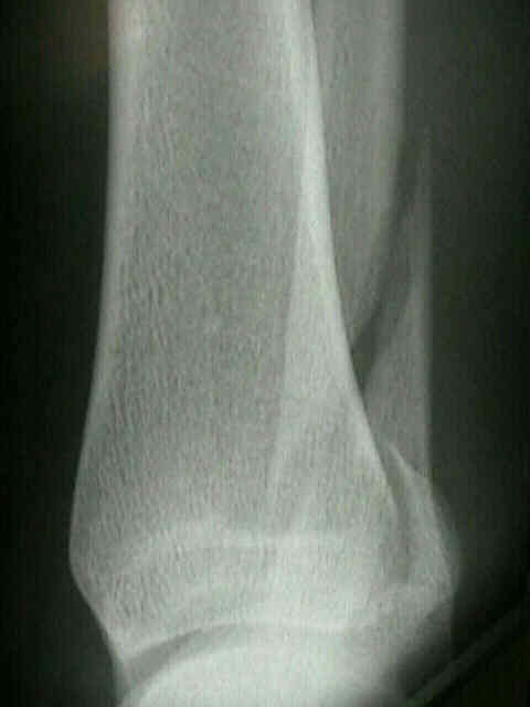
- rarely long posterior spike of distal fragment is comminuted;
- Fracture Characteristics:
- w/ supination external rotation frx, spiral oblique frx usually begins in almost transverse plane distally on anterior surface of the fibula at or just above level of plafond;
- it spirals externally, w/ frx exiting proximally on its posterior surface;
- hence, look for posterior spike;
- malleolar fragment carries the lateral attachment of ATLF
- this structure can often be a guide to reduction;

- Technique:
- fracture is distracted with longitudinal distraction and inversion of the foot opening the fracture site.
- fracture hematoma is curetted free from the bone ends.
- #15 blade was used to remove periosteum from edges of fracture site.
- reduction is obtained showing anatomic interdigitation of fracture fragments;
- reduce & internally fix lateral malleolus or fibular frx before fixing medial malleolus component;
- expose fracture & anterior surface of fibula proximal to it, explore joint, using an intra-articular angled retractor anteriorly;
- distal fibula is grasped with pointed reduction forceps & teased into position;
- simultaneous control of proximal fibular fragment w/ bone aids reduction;
- small, pointed or lobster claw reduction forceps is used to oppose frx as proximal and distal pieces are realigned;
- a useful technique to hold the reduction, involves insertion of one or two K wires across the frx site;
- following this, reduction clamps can be applied to facilitate insertion of a lag screw;
- K wires will have to be removed prior to lag screw insertion;
- unless extensively comminuted, posterior spike can guide restoration of length and rotational alignment;
- it may be repositioned first, & held in place while reduction is completed;
- once reduction is achieved, no talar tilt should remain;
- fixation of fibular in shortened or rotated position will often cause rapid dissolution of the ankle joint;
- usual reason for persistent valgus talar tilt is comminuted fibular fracture in which proper length has not be restored