

- See:
- Types of Loosening:
- Exam for THR Loosening:
- Grading of Cement Technique: (Barrack, et al. (1992) and Mulroy, et al. (1995))
- Grade A: meduallary canal completely filled w/ cement (white out).
- Grade B: a slight radiolucency exists at the bone cement interface.
- Grade C: a radiolucency of more than 50% at the bone cement interface.
- Grade D: a radiolucency involving more than 100% of the interface between bone and cement in any projection, including absence of
cement distal to the stem tip;
- as noted by Mulroy, et al., a femoral cement mantle less than 1 mm and defects in the cement mantle are associated with early
loosening;
- Improved cementing techniques and femoral component loosening in young patients with hip arthroplasty. A 12-year radiographic review.
- Total hip arthroplasty with use of so-called second-generation cementing techniques. A fifteen-year-average follow-up study.
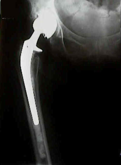
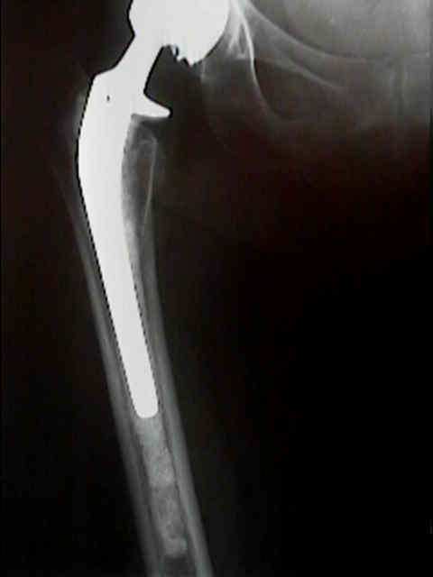
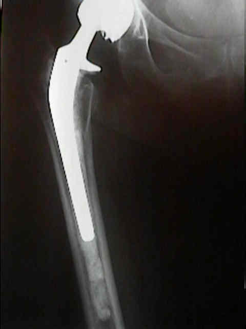
- Radiographic Stem Loosening:
- note that radiolucent lines are commonly found in the lateral and anterior aspects of the proximal femur, since these areas can be difficult to
visualize and difficult to pressurize;
- in the proximal 1 cm of the femur, linear radiolucencies less than 2 mm in width should not be considered to be indicative of
loosening;
- definite loosening:
- stem fracture
- cement fracture;
- cement-prosthesis lucency: radiolucency at the cement component interface greater than 1 mm in width;
- is a definite sign of loosening if a new radiolucent line of any size apprears at the cement prosthesis interface which was not
present on initial postoperative radiographs;
- cement debonding between the prosthesis and the cement in zone 1 (anterolateral segment), may not necessarily correlate with
poor clinical results;
- changes in stem position:
- pistoning:
- medial midstem pivot:
- calcar pivot:
- subsidence;
- distal pivot:
- probable loosening:
- radiolucent lines: continuous radiolucency at bone cement interface;
- typically these radiolucent lines will be surrounded by lines of increased density;
- endosteal cavitation (linear osteolysis and focal osteolysis) are also suggestive of femoral loosening;
- possible loosening:
- radiolucent lines at cement bone interface from 50-100% of the total bone cement interface;
- Technical Failures Causing Loosening:
- as noted by Kobayashi, et al (1997)., a "stove pipe" femur is a strong risk factor for radiographic loosening in cemented femoral components;
- Assessment of cement column:
Factors affecting aseptic failure of fixation after primary Charnley total hip arthroplasty.
Femoral component loosening using contemporary techniques of femoral cement fixation.