- Roberts View:
- true AP view described by Roberts:
- take AP view of thumb w/ forearm in maximal pronation & dorsum of thumb resting on the x-ray cassette;
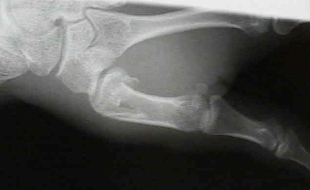
- True Lateral of CMC:
- obtained w/ forearm flat on table, hand pronated approx 20 deg w/thumb flat on cassette, & x-ray tube angled 10 deg from vertical in distal to proximal projection;
- allows evaluation of metacarpal displacement;
- estimates of the size and position of the volar fragment
- estimates gap between fragment & metacarpal base;
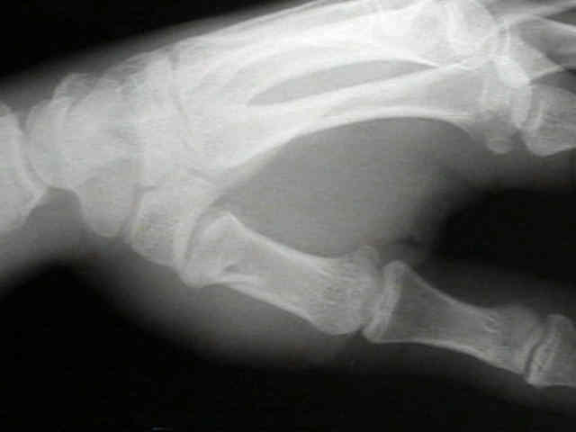
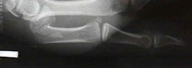
- Case Example:
- 20-year-old male who sustained extra-articular basal frx of thumb;
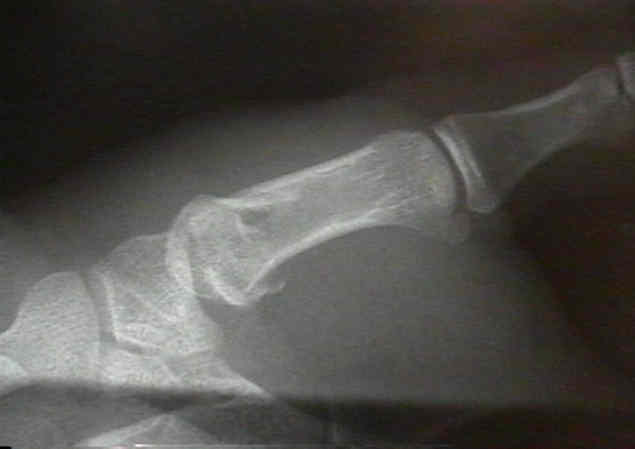
- on AP and Oblique views the displacement is not impressive;
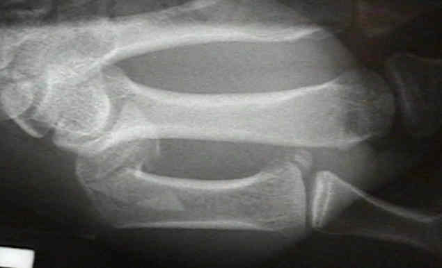
- on a true lateral view, however, the indication for surgery is apparent