- Discussion:
- evaluation of the painful hip replacement and exam for THR loosening:
- heterotopic ossification:
- post op AP & lateral x-rays should include entire length of stem & cement mass;
- femur and cement column are inspected carefully and compared w/ previous films for changes indicating component loosening, stem failure, trochanteric
problems, or infection;
- Radiographic Views: (bone scans for THR)
- frog leg lateral: gives best lateral of prox portion of femoral component;
- cross table lateral:
- for evaluation of position of acetabular component (anteversion) & status of post bone stock in posterior column & neck of ilium;
- weight bearing and non wt bearing views:
- may detect implant loosening;
- wt bearing view my demonstrate early polyethylene wear;
- push pull views: to evaluate for loosening of components;
- same as AP view of hip w/ examiner providing distraction and compression to the hip through the femur;
- references:
- A Systematic Approach to the Plain Radiographic Evaluation of the Young Adult Hip
- Leg Length and Offset: (see femoral offset)
- leg length descrepancy: (see intraoperative leg length determination)
- measured by noting the realative positions of the lesser trochanters from a line drawn tangential to the ischium;
- as noted by Ranawat (1999), 87% of patients had leg lengths within 5 mm of each other;
- note that with an increased offset femoral component, the gluteus medius may be tight which causes a pelvic tilt, and gives an
apparent leg length inequality;
- as the gluteus medius stretches out over several months, the apparent leg length inquality decreases toward normal;
- reference:
- Hip arthroplasty: postoperative management problems. The pants too short, the leg too long.
- Clinical significance of leg-length inequality after total hip arthroplasty.
- Functional leg-length inequality following total hip arthroplasty.
- Surgical Treatment of Limb-Length Discrepancy Following Total Hip Arthroplasty.
- Leg-length Inequalities Following THA Based on Surgical Technique
- Limb-length Discrepancy After Total Hip Arthroplasty
- Limb-length Discrepancy After Total Hip Arthroplasty: Novel Treatment and Proposed Algorithm for Care
- Total hip arthroplasty: leg length inequality impairs functional outcomes and patient satisfaction
- A radiographic comparison of femoral offset after cemented and cementless total hip arthroplasty.
- radiology of offset:
- horizontal and vertical offset depends on the the amount of acetabular reaming, femoral neck cut, inaddition to the modular components in
both the acetabular cup and femoral stem;
- the easiest way to judge offset is to compare Shenton's line of the opposite (normal hip) to the operative hip;
- a break in Shenton's line along with decreased area under the curve indicate decreased offset;
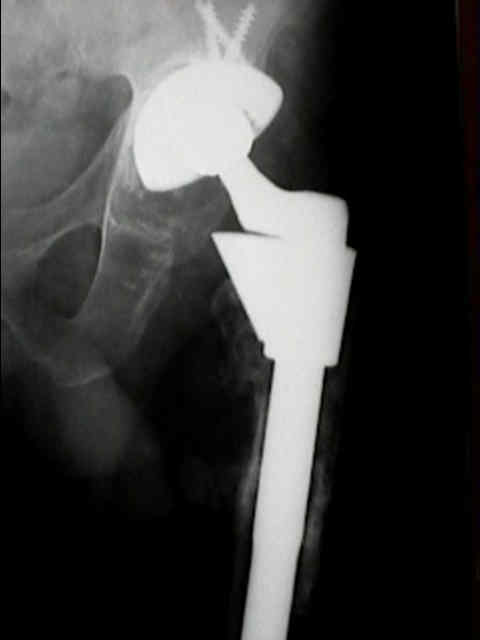
- Radiology of Acetabular Component:
- note that plain radiographs may be more accurate for identifying femoral component loosening than for acetabular loosening;
- radiographic evaluation of cup position:
- Judet views:
- cross table lateral:
- for evaluation of position of acetabular component (anteversion) & status of post bone stock in posterior column & neck of ilium;
- Lowenstein lateral radiograph:
- provides a lateral view of the acetabular subchondral bone and the cup after implantation
- modified Lowenstein lateral radiograph is similar to an oblique radiograph of the pelvis;
- patient is turned onto the affected hip at least 45° and as much as necessary to allow the lower limb to be in abduction and
external rotation and to be flat on the x-ray table;
- polyethylene wear (need to rule out osteolysis w/ annual radiographs);
- acetabular component loosening
- where as patients w/ loose femoral components will often complain of pain, in contrast, patients w/ loose acetabular components may be asymptomatic;
- asymptomatic patients w/ radiographic evidence of loosening need to be followed for implant migration and loss of bone stock;
- some surgeons will recommend revision for radiographic loosening even if patients have no symptoms;
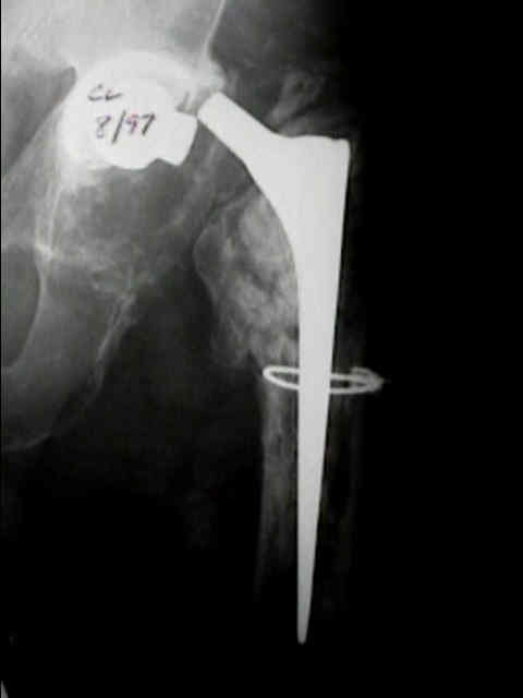
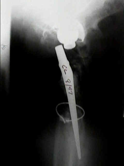
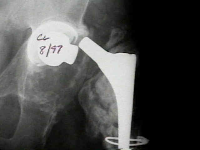
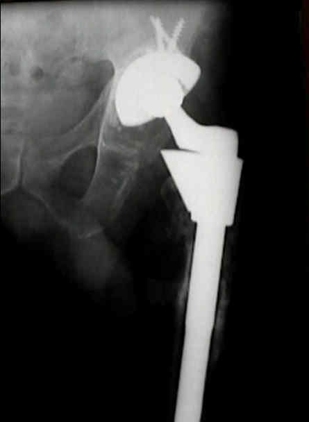
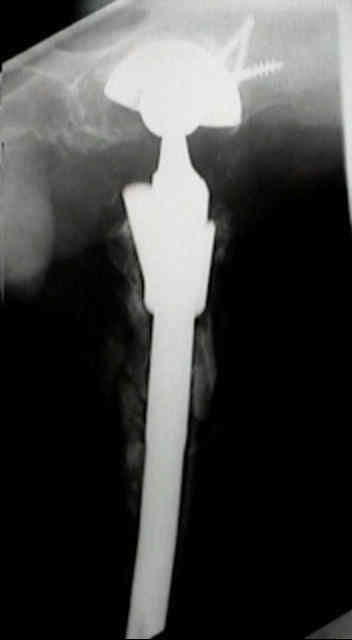
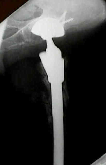
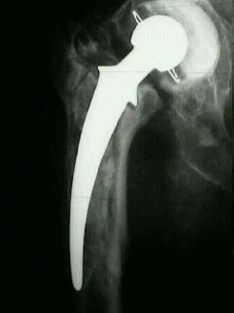
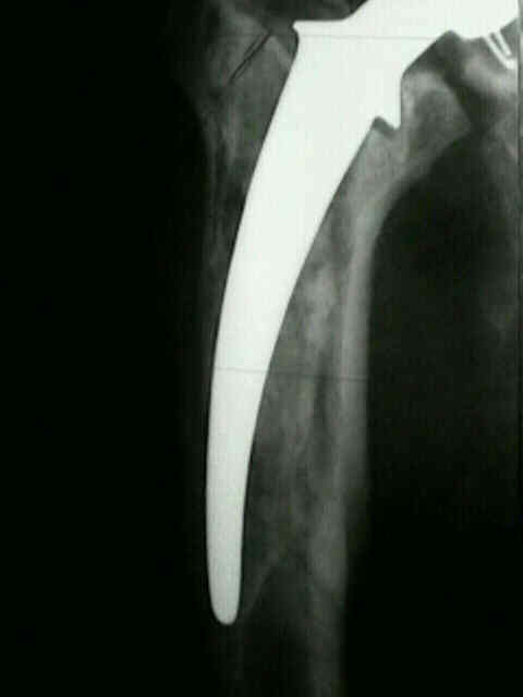
- Radiology of Femoroal Component: (post op x-rays for thr)
- post op ap & lateral x-rays should include entire length of stem & cement mass;
- yearly radiographs need to be taken to look for progressive osteolysis;
- radiographic views:
- frog leg lateral: gives best lateral of prox portion of femoral component;
- stress views:
- may detect implant loosening;
- weight bearing and non wt bearing views:
- push pull views: to evaluate for loosening of components;
- same as ap view of hip w/ examiner providing distraction and compression to the hip through the femur;
- implant migration (indicates loosening);
- pistoning / subsidence:
- medial midstem pivot
- calcar pivot: (distal toggle)
- bending cantilever (distal pivot)
- cemented femoral component:
- femur and cement column are inspected carefully and compared w/ previous films for changes indicating component loosening, stem
failure, trochanteric problems, or infection;
- press fit femoral component:
- end of stem pain is usually present from time of surgery, tends to improve during 1st year, but may remain constant thereafter;
- although bone scans may help, many noncemented THR, esp long stems, may show some increase in activity;
- divergent radiolucent lines in area of ingrowth indicate loosening;
- varus / valgus positioning:
- traditionally, varus positioning has been thought to lead to premature loosening;
- in the report by Sochart and Porter (1997), however, neither varus or valgus stem position appeared to be associated w/ premature
stem loosening (w/ average of 20 years follow up)
The histology of the radiolucent line.
Quality assessment of early versus late post-operative radiographs in joint replacement surgery.
The Internet Journal of Orthopedic Surgery. 2008 Volume 10 Number 2. Z.A. Jibri, S.R. Fernando, S.B. Mirza, M.M. Glasgow.
We assessed the post-operative radiographs of 87 patients who had total hip (46 patients) or total knee arthroplasty (41 patients).
This study has also demonstrated that the late post-operative radiographs following THR are of better quality than the early ones. These early radiographs were of poor quality and we question their role as a baseline for further examinations.
..... If the first postoperative radiographs were taken in the follow up clinic, this would result in a better quality radiograph, in a more comfortable patient easily positioned by the radiographer. It will reduce the pressure on the radiology services as the patient would not need to have the X-ray during the hospital stay. It will also help in the logistics with in the hospitals as the patient would normally walk to the radiology department in the late postoperative period and would not need transport.
In conclusion, this study has demonstrated that there is a significant difference in the quality of post-operative radiographs in favour of the late films.
TOTAL HIP REPLACEMENT – ARE CHECK RADIOGRAPHS REQUIRED?
J Bone Joint Surg Br 2006 vol. 88-B no. SUPP II 249. JR Crawford, I Syed, M Babatope and GS Keene
Recovery room radiographs after total hip arthroplasty: tradition vs utility?
J Arthroplasty. 2012 Jun;27(6):1051-6. doi: 10.1016/j.arth.2011.12.020. Epub 2012 Feb 2.
Ndu A, Jegede K, Bohl DD, Keggi K, Grauer JN.
In a review of 632 consecutive recovery room series, Findings suggest that the single routine inpatient series should be taken in the radiology suite, rather than in the recovery room.
Routine recovery room radiographs after total hip arthroplasty: ineffective for screening and unsuitable as baseline for longitudinal follow-up evaluation.
J Arthroplasty. 2004 Apr;19(3):313-7. Mulhall KJ, Masterson E, Burke TE.
retrospectively analysed 2,065 consecutive hip arthroplasty patients... found a 0.1% rate of radiologic diagnosis of dislocation in the population screened. In 100 patients randomly selected for comparison, the image quality in the recovery room radiographs was significantly inferior to standardized departmental radiographs (P<.001), with further significant differences between cup version (P<.001), and stem alignment assessments (P=.002). With such poor information and diagnostic yield for follow-up and screening, these investigations should only be performed when clinically indicated.
Postoperative radiographs following hip fracture surgery. Do they influence patient management?
Int J Clin Pract. 2007 Mar;61(3):421-4. Chakravarthy J, Mangat K, Qureshi A, Porter K.
conduct a national audit on current UK practice regarding the use of check radiographs following hip fracture surgery. Retrospective case note review of all patients undergoing hip fracture surgery at our hospital, from 2002 to 2004, was performed. Patients undergoing revision surgery in the same admission were identified to determine whether check radiograph influenced the decision. Subsequently a postal performa was sent to 450 randomly chosen UK Orthopaedic Consultants. The performa was designed to determine practice relating to postoperative radiographs. It also attempted to determine whether postoperative radiographs (when requested) influenced the subsequent clinical management of the patient. A total of 1265 hip fractures treated surgically were reviewed locally. .....The study highlights the lack of national consensus on the use of postoperative radiographs. We recommend that following DHS/DCS fixation and CS fixation, the use of postoperative radiographs should only be undertaken when clinically indicated. Postoperative radiographs following hip hemiarthroplasty should only be undertaken if there are operative concerns or postoperative complications.
Check radiography after fixation of hip fractures: is it necessary?
J R Coll Surg Edinb. 2000 Dec;45(6):398-9. Mohanty K, Gupta SK, Evans RM.
requesting routine post-operative check radiographs for these fractures are still a common practice...
We suggest that routine post-operative radiographs after femoral neck fracture fixation are unnecessary unless there is some clinical indication. This has significant implications in relation to patient discomfort, radiation exposure and cost-effectiveness.