

- Methods to Etimate Growth Potenital:
- Growth Plate Anatomy
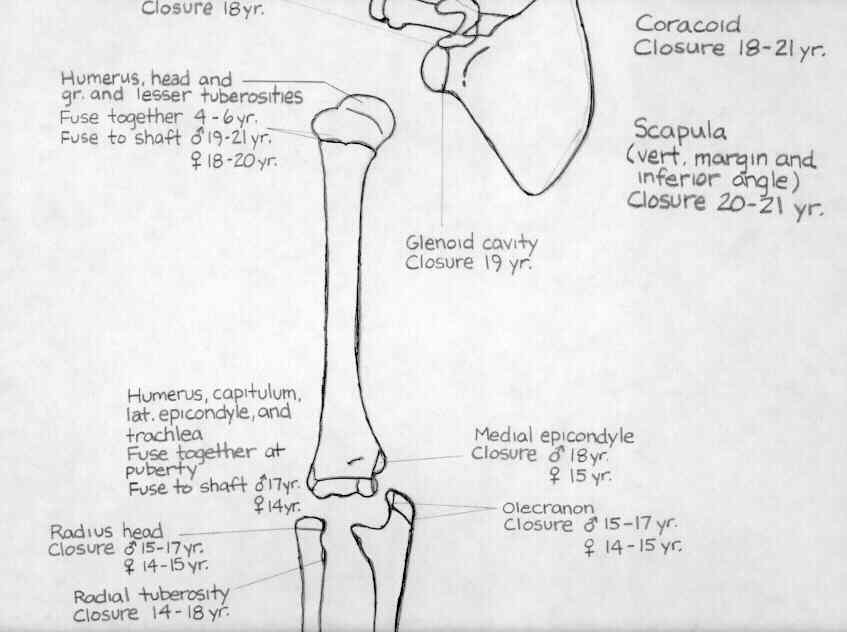
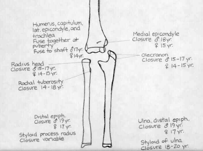
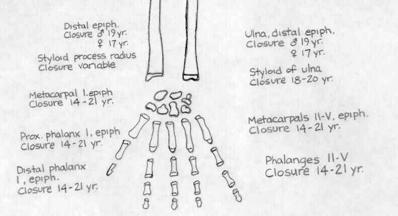
- Appearance of Ossification Centers:
- skeleton incompletely ossified at birth, & portions such as carpals, patella, navicular, cuneiforms, will radiographically appear entirely cartilaginous;
- ossification centers will enlarge in all directions, until it becomes bounded on metaphyseal side by the epiphyseal plate and on all other sides by articular cartilage;
- at this point, the nuceus grows only on the sides adjacent to cartilage;
- primary ossification centers at 8 weeks gestation in both radius and ulna
- secondary ossification centers arise between birth and the 20th year;
- fusions of these secondary centers w/ main bones usually occur in late adolescence;
- specific ossification centers make their appearance in a specific order:
- ossification centers in calcaneus and talus appear in the sixth to eighth fetal month (or earlier);
- distal femoral epiphysis & proximal tibial epiphysis is present at birth;
- cuboid appears at the time of or soon after birth;
- proximal femoral epiphysis appears between 2 - 8 months following birth;
- distal epiphyses of radius appears radiographically at age 1 - often from two centers
- ossification center of the greater trochanter appears at 2 yrs and fuses to metaphysis at 16 years;
- radial head epiphysis is not present until after 3 years of age (may occur between 5-7 years);
- distal ulnar epiphysis appears at age 5;
- ossific nucleus of the pisiform appears at about 6 to 8 years of age
- olecranon appears between 9-10 years;
- Chronologic Age by Risser's Sign:
- Girls Boys
- Risser Age Risser Age
1 13.8 1 15.2
2 14.3 2 15.2
3 14.7 3 16.3
4 16.0 4 16.3
5 16.1 5 18.0
- Approximate Contributions to Growth: (until age 13 for females and age 15 for males)
- proximal femur = 1/8 in./year
- distal femur = 3/8 in./year
- proximal tibia = 1/4 in./year
- distal tibia = 3/16 in./year
- total adult ht = ht. at age 2 x 2
- growth ceases: 15 - 15 1/2 yrs for girls, 17 - 17 1/2 yrs for boys
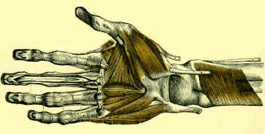
- hand of a 10 year old male;
Accuracy of the Sauvegrain Method in Determining Skeletal Age During Puberty.