

- Radiology:

- SH Type I Frxs:
- separation of epiphysis occurs thru hypertrophying layer of cartilage cells;
- proliferating cells are intact, the epiphysis continues to grow;
- if nutrient artery is intact healing occurs in 3 weeks;
- frx is most common in distal phalanx, uncommon in middle and proximal digits;
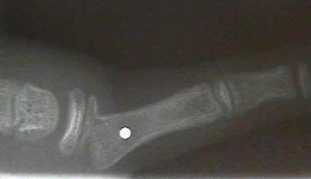
- type I epiphyseal frx / separation of distal phalanx;
- phalanx is usually flexed & displaced laterally;
- epiphysis remains extended relative to phalanx;
- closed reduction: traction w/ lateral & vertical pressure on joint & phalanx;
- finger is immobilized w/ external splint or smooth K wire inserted thru distal phalanx, epiphysis, and DIP joint;


- Phalangeal Neck Frx:
- initial displacement determines how frx is treated;
- usually there is volar angulation at the frx site;
- minimally displaced fractures are reduced and splinted for 3-4 weeks, which is then followed w/ 1-2 weeks of buddy taping;
- displaced fractures usually will require K wire fixation - pins are left in place for 3 weeks, followed by buddy taping;
- there is little potential for remodeling in this fracture;
- Accetable Reduction:
- AP angulation:
- some remodeling may occur in both metacarpal and phalangeal frx;
- lateral angulation: will not remodel;
- rotary deformities: will not remodel;
- use finger nails to help judge rotation;
- Treatment Principals:
- most pediatric phalangeal frx can be managed w/ gentle reduction, and splinting for 3 weeks;
- splinting involves gutter splint and usually incorporation of one un-injured digit (which assists w/ control of rotation)
Fractures about the interphalangeal joints in children.
Subcondylar fossa reconstruction for malunion of fractures of the proximal phalanx in children.
Fractures of the phalanges of the hand in children.
Remodelling of a displaced phalangeal neck fracture.
Physeal and periphyseal injuries of the hand. Patterns of injury and results of treatment.
Percutaneous Reduction and Fixation of Displaced Phalangeal Neck Fractures in Children