

- osteomyelitis occurs most frequently in the long bones of lower extremities, & to a lesser extent the upper extremities;
- hematogenous osteomyelitis
- pediatric bone circulation: tortuous course of nutrient vessels in bone causes bacteria to be trapped in the metaphysis;
- epiphyseal plate prevents infection from entering the joint space in older children but not in neonates;
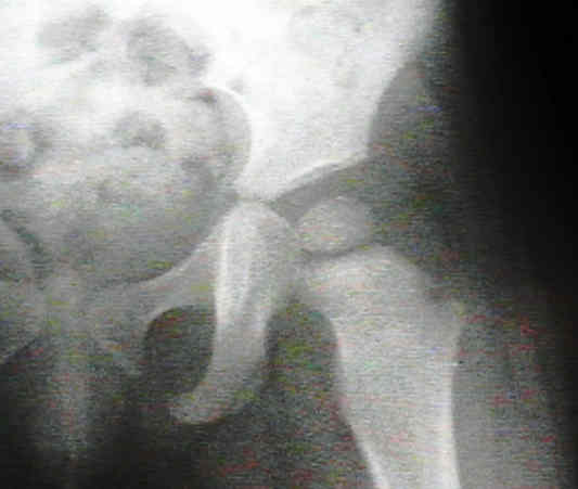
- joint infection secondary to osteomyelitis may occur in shoulder, radial head and the hip as a result of synovial membrane inserting
distally to epiphysis, allowing bacteria to spread directly from metaphysis to joint space;
- infecting organism may reach the joint in three ways:
- hematogenous seeding of synovial membrane
- extension from an adjacent focus of osteomyelitis
- direct implantation from a penetrating wound;
- bacteriology:
- ages 6 mo to 2 yrs
- ages over 2 yrs
- predisposing conditions:
- chicken pox: streptococcus
- ear infection: h. influenza or streptococcus;
- sickle cell: salmonella;
- meningitis:
- references: The changing epidemiology of osteomyelitis in children.
- Work Up:
- before aspiration, consider whether an MRI is indicated (you do not want to aspirate and EOG without a prestage MRI);
- aspiration of site:
- in order to recover causitive organisms & to determine whether an abscess is present, which wound require surgical drainage;
- drilling is used to decompress known proximal femoral osteomyelitis that has not already decompressed into the hip joint and to obtain a Gram's stain and culture material in cases of possible primary bone infection with sympathetic hip effusions;
- in 1974, Kemp and Lloyd-Roberts noted several cases of osteonecrosis after proximal femoral osteomyelitis w/o apparent hip sepsis;
- based on this, they recommended drilling all proximal femoral osteomyelitis;
- reference: Avascular necrosis of the capital epiphysis following osteomyelitis of the proximal femoral metaphysis
- Treatment:
- antibiotics:
- surgical debridement and vancomycin Ca sulfate
- references:
- Treatment of chronic osteomyelitis in children resistant to previous therapy.
- Complications:
- deep venous thrombosis
- Deep Venous Thrombosis Associated with Osteomyelitis in Children
- Upper-Extremity Deep Venous Thrombosis Associated with Proximal Humeral Osteomyelitis in a Child. A Case Report
- Deep venous thrombosis associated with acute hematogenous osteomyelitis in children.
- Venous thrombosis and thromboembolism in children with osteomyelitis.
References:
Pediatric Humeral Osteomyelitis
Osteomyelitis in infants and children. A review of 163 cases.
Primary subacute epiphyseal osteomyelitis.
Diaphyseal primary subacute osteomyelitis in children.
Osteomyelitis of the calcaneus in children.
Occult epiphyseal bone abscess: lessons for the unwary.
Hematogenous osteomyelitis of the wrist in children.
Osteomyelitis of the pelvis in children.
The three syndromes of iliac osteomyelitis in children.
Pathologic Fractures in Children with Acute Staphylococcus aureus Osteomyelitis