

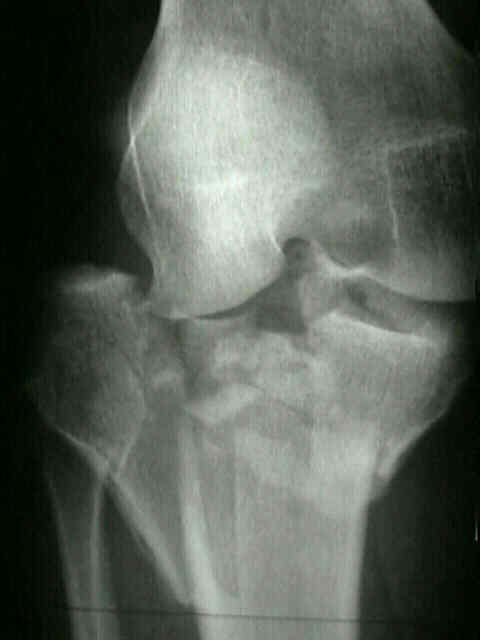
- Technique: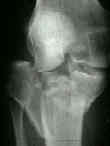
- stratedgy involves, performing articular surface reduction, and then to secure proximal portion to tibial metaphysis
w/ use of lateral butress plate along w/ possible use of medially applied external fixation;
- application of lateral plate takes advantage of the better soft tissue envelop on the lateral side of the leg;
- supine position:
- tape a rolled sheet onto the table (as with a total knee replacement), inorder to allow the knee to remain hyperflexed during the procedure (when required);
- supine w/ leg flexed:
- leg can be positioned as for arthroscopy;
- a leg holder is applied to the proximal thigh, and the table is broken to allow the knee to flex past 90 deg;
- the opposite leg is then held in a GYN leg holder;
- this technique allows varus to be applied to the knee which improves fracture exposure;
- indirect reduction:
- preceeds open reduction;
- apply femoral distractor to medial aspect of knee;
- exposure:
- use longitudinal incision, rather than curved hockey stick;
- this approach will not interfere w/ a TKR incision if one is needed in the future;
- parapatellar incision is made starting above the superior pole of the patella, and is brought down on either medial or the lateral side
of the patella, depending on the location of the major fracture line;
- incision continues for about 3-4 cm below the lowest extent of the frx;
- patellar tendon Z-plasty:
- contra-indicated if there is concomitant tibial tubercle frx;
- for improved exposure, consider cutting the patella in a "Z pattern" (longitudinal split w/ half attach to tubercle and half attached to patella)
with repair of the tendon with heavy suture at the end of the case;
- deep dissection depends on the location of the major frx line
- see exposure of: lateral and medial plateau;
- anterior meniscal detachment:
- see: lateral meniscus;
- just below the joint line, make a horizontal inframeniscal incision, which extends from a point anterior to
the frx fragment to a point posterior to the frx fragment;
- infra-meniscal incision needs to be large enough to allow the meniscus to be elevated and the frx to be
"booked open";
- sharply detach the anterior horn of menisci from its bony insertion;
- the anterior horn will later be repaired at the end of the case;
- this allows the surgeon to pull up on meniscus together w/ the attached capsule, for more optimal view of articular surface;
- meniscus is retracted proximally, and the joint inspected directly;
- if the arthrotomy is made above the meniscus, the meniscus will keep most of articular surface hidden from view;
- the knee is hyperflexed and internally rotated;
- meniscal re-attachment:
- two small drill holes are made at the origin of the anterior horn and are directed to exit medial to the patellar tendon;
- pass a suture thru the meniscus, and use a suture passer to thread the sutures thru the drill holes;
- optimize the expsure:
- the detached meniscus should be retracted proximally;
- the knee is hyperflexed and internally rotated;
- open reduction:
- split fracture fragments can be booked open to allow elevation of depressed fragments;
- in some cases, a window may have to be created to assist w/ articular elevation;
- bone graft may then be inserted into the window to support the elevated articular fragments;
- medial and lateral bone fragments are reduced under direct vision;
- percutaneously applied reduction forceps may assist w/ reduction;
- k wire fixation:
- k wires are inserted to maintain provisional fixation;
- take care that k wire position does not interfere with plate application;
- apply lateral butress plate;
- apply medial external fixator or consider circular wire fixation;
- medial plateau is usually much less comminuted than lateral plateau & medial plateau is more ammenable to fixation w/ screws than lateral plateau;
- because the lateral plateau is higher, one has to be careful with the insertion of proximal screws medially
from lateral side so as not to enter the medial joint space;
- locking plate fixation (see synthes)
- posteromedial incision (for secondary coronal plane fracture);
- plane between the semitendinosis and gastrocnemius
- ref: Posterior coronal plating of bicondylar tibial plateau fractures through posteromedial and anterolateral approaches in a healthy floating supine position.
- wound closure:
- menisci are repaired using nonabsorbable suture, w/ the repair beginning posteriorly and proceding anteriorly;
- posterior sutures are placed thru the iliotibial tract and the anterior sutures are placed thru the parapatellar retinaculum