

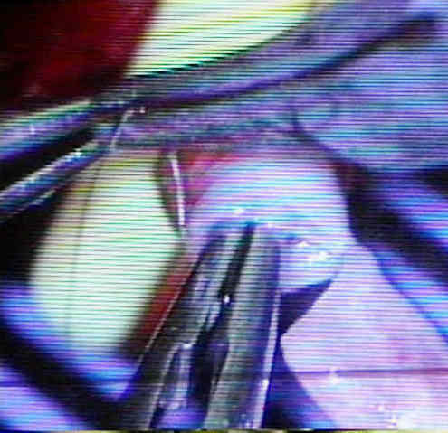

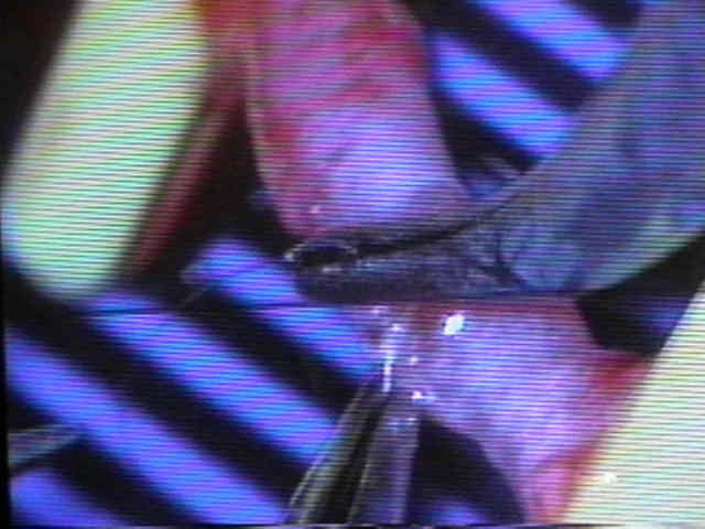
- Vessel Exposure: Exposure of the donor vessels is optimized with the use of a large self retaining retractor superior in the wound as well as the Charnley retractor. The microscope is brought in to perform the anastamosis. The vein is coupled first. This prevents any bleeding which could obscure the field if the artery was anastamosed first. The vein is held in position with a disposable plastic vessel approximator. We use a 3M coupler device for the vein only. This cuts about 20 minutes from the operating time compared to a hand sewn anastamosis. The coupler may be inadequate for the artery because of the thickness of the arterial wall. When the artery is stretched over the coupling device, intimal cracking has been observed. A suction micro-mat of contrasting colour is helpful in performing the microsurgery. The operation is easier if all sewing of the artery is done from the dorsum of the patient. The microscope is positioned such that the operator is looking about 30 degrees cephalad instead of perpendicular to the patient.
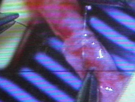
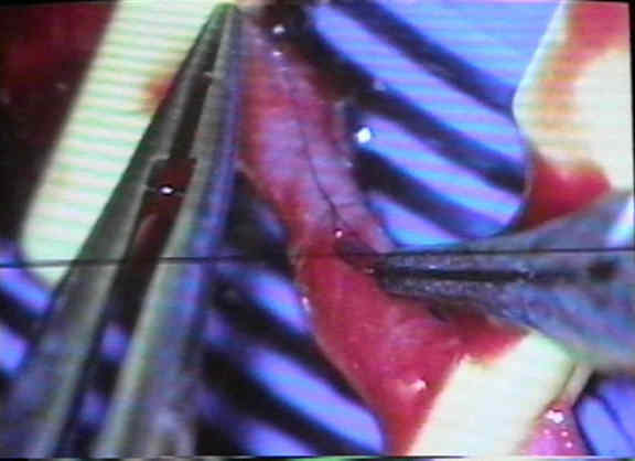
- Vessel Anastomosis: The artery is sewn with interrupted 8-0 nylon suture after confirming good proximal flow. The distal clamp is released first. The clamped areas of the vessel are massaged gently with the jewelers forceps to break up any microemboli. The vessels are irrigated with warm solution. The microscope is positioned over the fibular end, visible in the wound at the hip. There should be bleeding from the endosteal vessels. Often there can be blood welling from the haversian system of the bone. There are minimal problems if the anastamosis is done in a timely fashion. A clamp time of less than 15 minutes is optimum. If there is no bleeding or propagated pulse in the artery, then other measures have to be taken. The room is warmed and a papaverine solution is placed onto the pedicle. Young female patients have a predilection towards spasm and the room should be kept warmer in these patients. Their pedicle should be manipulated minimally, if possible. If there is still no transferred pulse and filling tests are negative, then the anastamosis has to be taken down to examine for a mechanical reason of decreased flow, such as a back wall suture. We have found that the majority of these cases are due to spasm and not a technical error. The spasm in the vessel can be broke with a dilator by gently passing it past the area of constriction. This increases the chance for later spasm but verifies that there is no mechanical impediment to flow. The artery is resutured and the wound closed. In earlier patients we had performed bone flow scans to see if there was blood flow in the fibula. The patients with spasm regained normal flow after closure.