


- See: Scaphoid Frx Menu:
- scaphoid nonunions:
- Discusssion:
- bone grafting may be indicated for scaphoid malunion or non union;
- malunion:
- indicators of scaphoid malunion: classic findings are a dorsal humpback deformity and DISI deformity;
- indications for bone grafting include radiographic deformity along with pain, weakness, and loss of motion;
- note that the proximal fragment may lie supinated, extended and radially deviated relative to the distal fragment;
- even small degrees of malunion will result in a significant loss of extension;
- Russe used a volar approach to the scaphoid, believing that it was less disruptive to the blood supply as well as allowing insertion
of graft from front to correct the angulatory deformity;
- contra-indications:
- operative treatment with Matti-Russe grafting does not reliably lead to union of proximal pole nonunion with AVN
- this technique is contra-indicated if proximal pole is avascular
- vascularity of the scaphoid cannot always be predicted by radiographs;
- best way to determine vascularity is to look for punctate bleeding at the time of surgery;
- relative contra-indication is a severe DISI deformity, since adequate correction of deformity may not be achieved;
- Surgical Approach:
- when performing a volar approach, radioscaphocapitate ligament and palmar radiolunate-triquetral ligament must be partially or
totally divided;
- if inadequately repaired, the natural tendency of the lunate to extend and the scaphoid to flex under axial compression may
lead to a DISI deformity;
- radial styloidectomy:
- radial styloidectomy may help decrease postoperative radial sided tenderness;
- may be performed through a separate posterolateral incision;
- attempt to preserve the radiocapitate ligament;
- pseudarthrosis is resected back to healthy bone where possible;
- this is best done using small osteotome to square off both frx faces;

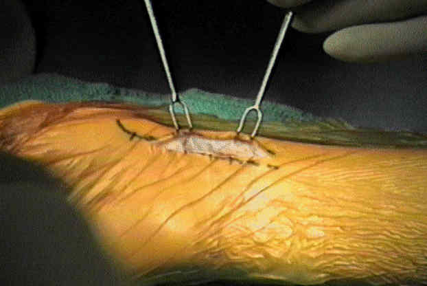
- Bone Graft:
- bone graft must be taken from iliac crest;
- 1.5-cm block of corticocancellous bone is removed from outer table of iliac crest after apex has been hinged off;
- graft is shaped to fill defect left following resection of pseudarthrosis & correction of deformity by dorsiflexion of wrist;
- consider applying the inner wall of the ileal graft toward the capitate for better congruence;
- cortical component should lie flush w/ anterior surface of scaphoid to provide mechanical stability on compression;
- whenever possible, a soft-tissue hinge should be preserved posteriorly to retain the graft and provide extra stability;
- alternative graft technique:
- harvest two cortico-cancellous grafts, which are inserted into the excavated scaphoid, w/ the cancellous sides facing internally;
- Hardware Insertion:
- cannulated screw fixation:
- herbert screw insertion technique:
- wherever possible, jig should be applied in such a way that screw will be perpendicular to fracture.
- if obliquity of frx line makes this impossible, supplementary K wire fixation should be used to prevent shearing displacement
under compression;
- drill guide is placed across the distal pole, and the blade is placed across the proximal pole;
- frx site is compressed by jig prior to screw placement;
- jig is compressed as tightly as possible after any remaining gaps between the graft and frx faces have been filled with
cancellous bone chips;
- measure screw size, and insert screw below surface of scaphoid
Fixation and Grafting After Limited Debridement of Scaphoid Nonunions
Hybrid Russe Procedure for Scaphoid Waist Fracture Nonunion With Deformity
Long-term results after Russe bone-grafting: the effect of malunion of the scaphoid.
Scaphoid nonunion treated with the Matti-Russe technique. Long-term results.
The effect of avascular necrosis on Russe bone grafting for scaphoid nonunion.
Corrective Osteotomy for scaphoid malunion: technique and long term follow up evaluation.
Scaphoid Waist Nonunion With Humpback Deformity Treated Without Structural Bone Graft