

- See: Lisfranc's Frx
- Anatomy:
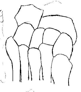
- proximal end of the second metatarsal is tightly recessed between first and third cuneiforms;
- this mortise configuration effectively locks entire tarsometarsal complex, preventing medial or lateral translation;
- no significant dislocation of metatarsals or cuneiforms can occur unless this bone is disrupted;
- for this reason, pure transmetatarsaltarsal dislocations rarely occur, & rather involve frxs through or around the second metatarsal base;
- there are no ligaments binding together the first and second metatarsals;
- this creates a relative weakness between 1st & other metatarsals;
- the main stabilizer of the 1-2 intermetatarsal joint is Lisfranc's ligament;
- Lisfranc's ligament:
- strong oblique ligament which extends from plantar-lateral aspect of medial cuneiform, passes in front of the intercuneiform ligament, and inserts into the plantar-medial of second metatarsal;
- variations:
- in about 20% of patients, two separate bands of the ligament are present (dorsal and plantar);
- in patients with two separate ligamentous bands, partial ligament injuries are possible;
- function:
- acts to connect lateral metatarsals to the medial cuneiform;
- reinforces bony stability of base of the 2nd metatarsal between medial and lateral cuneiforms;
- dorsalis pedis artery:
- crosses Lisfranc's joint & dives deep between bases of the first and second metatarsals to form plantar arterial arch, making it susceptible to damage at time of injury or open reduction;
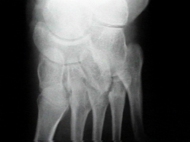
- Radiographs:
- lateral view:
- metatarsal is never more dorsal than its respective tarsal bone but, on occassion, may be slightly plantar to tarsal bone;
- AP view:
- medial borders of 2nd metatarsal base & medial border of middle cuneiform, normally form a straight, unbroken line;
- disruption of this line indicatives unstable TMT injury;
- oblique view:
- allows evaluation of the lateral midfoot;
- medial border of 4th metatarsal base & medial border of cuboid, normally form a straight unbroken line;
- Equivocal Injury:
- fractures of the base of the 2nd metatarsal should always rainse suspicions of tarsometatarsal injury;
- any comminution or diastasis between the medial cunnieform and 2nd metatarsal indicates functional disruption of Lisfranc's ligamentous complex;
- wt bearing AP:
- w/ questionable injury, consider wt bearing AP view to assess 1-2 interval;
- diastasis of the 2nd metatarsal-medial cuneiform articulation, or widening of the first 1-2 intermetatarsal interval greater than 2 mm (compared to the opposite foot) indicates subluxation and warrents closed reduction and percutaneous scew fixation;
- Potter, et al (1998) noted that normal separation of 1-5 mm may be found between the metatarsals, and therefore it is vital to compare the injured foot to the uninjured foot;
- if standing AP is unacceptable to the patient then consider CT scan;
- abudction stress AP:
- in the study by Coss HS, et al (1998), cadavers had ligamentous sectioning and then underwent abduction stress AP x-rays;
- motivation for the study is the observation that w/ Lisfranc strain, abduction stress will move the forefoot laterally;
- in a control population a line tangential to the navicular and medial cuneiform (medial column line) intersected the base of the first metatarsal (even with abduction stress);
- in cadavers w/ ligamentous sectioning and applied abduction stress, the medial column line falls medial to the metatarsal;
- the authors also noted that the abduction stress AP needs to be taken w/o pronation or supination;
- of note, these authors noted that cadavers w/ ligamentous sectioning, did not show more than 1.5 mm of widening w/ simulated wt bearing;
- MRI:
- use of MRI in Lisfranc fractures to evaluate the Lisfranc ligament was studied by HG Potter MD et al. 1998.
- axial views were used to visualize the Lisfranc ligament;
- these authors noted that all patients with complete ligament tears had at least 2 mm or more displacement between the second metatarsal and medial cuneiform (compared to the opposite side);
- they suggest that an MRI be ordered when there is equivocal widening and likewise that an MRI not be ordered when the diastasis is greater than 2 mm (since ligament disruption is most likely present)
Magnetic resonance imaging of the Lisfranc ligament of the foot.